Figures & data
Figure 1. 1 H-NMR spectrum of the PLA (A), HA (B) and HA–ADH–PLA (C).
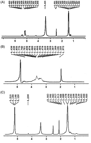
Figure 2. Infrared spectra of HA, HA–ADH and HA–ADH–PLA.
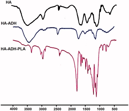
Figure 3. Dynamic light scattering (DLS) (A) and Field emission scanning electron microscopy (FE-SEM) (B) characterization of curcumin-encapsulated HA–PLA NPs.
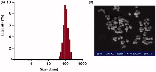
Figure 4. Drug release profiles of curcumin-encapsulated HA–PLA NPs in PBS at pH 4.4 and 7.4. The data are presented as mean ± SD (n = 3).
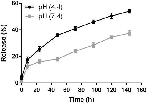
Figure 5. Cellular uptake of free curcumin (A) and nanoformulated curcumin (B) after 24 h of treatment using confocal microscopy (DAPI-blue light, curcumin-green light).
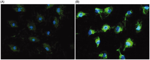
Figure 6. Effect of different concentrations of free curcumin and nanoformulated curcumin for 24 h on peritoneal macrophage viability. Macrophage viability was determined by MTT assay. Data were expressed as the mean ± SD of 3 independent experiments. *p < .05 compared to 15 and 30 μM nano-curcumin.
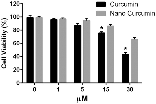
Figure 7. In vitro polarization study. iNOS-2 (M1 marker) and Arg-1 (M2 marker) in M1 peritoneal macrophages treated with free curcumin and curcumin-encapsulated HA–PLA NPs in different concentration for 24 h. *p < .05 compared to M1 macrophages that were attained by stimulating macrophages with LPS/IFN-γ, n = 3.
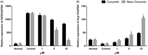
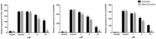
Figure 8. In vitro anti-inflammatory study in peritoneal macrophages. expression of pro-inflammatory cytokines, TNF-α, IL-1β, and IL-6 mRNA level at 24 h after treatment of macrophages with free curcumin and curcumin-encapsulated HA–PLA NPs, followed by stimulation with LPS/IFN-γ. qPCR was applied to measure mRNA levels. *p < .05 versus LPS/IFN-γ-treated group, n = 3.