Figures & data
Table 1. Characteristics of various formulations of nanoparticles (n = 3, mean ± SD).
Figure 1. Particles size distribution and transmitted electronic microscopy (TEM) of CUR-NPs (A, D), DOX-NPs (B, E) and CURDOX-NPs (C, F).
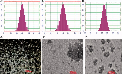
Figure 2. In vitro release profiles of DOX (A) and CUR (B) from nanoparticle formulation (CURDOX-NPs) or solution formulations (DOX-S or CUR-S) in pH7.4 PBS or pH5.0 acetate buffer.
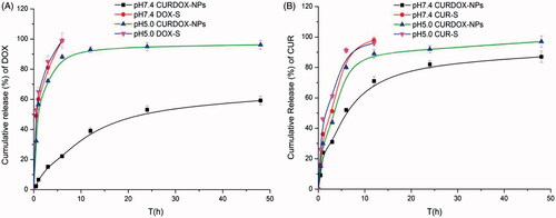
Figure 3. Cytotoxicity of the different formulation against MCF-7/ADR after incubation of 24 h (A) and 48 h (B), and (C) the fluorescent distribution of DOX in MCF-7/ADR cells after treatment with different formulations for 4 h at 37 °C.
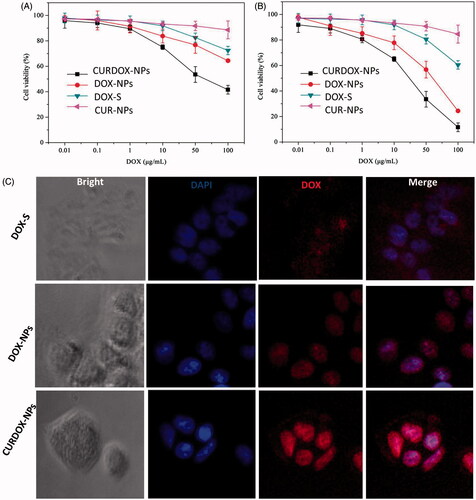
Figure 4. (A) microscopic images and (B) the relative spheroid volume of MCF-7/ADR spheroid after treatment with different formulations, and (C) the percentage of CD44+/CD24− cells in MCF-7/ADR mammospheres after 3 days of treatment with various formulations. The dissociated MCF-7/ADR mammosphere cells were stained with anti-CD44-FITC and anti-CD24-PE antibodies.
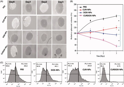
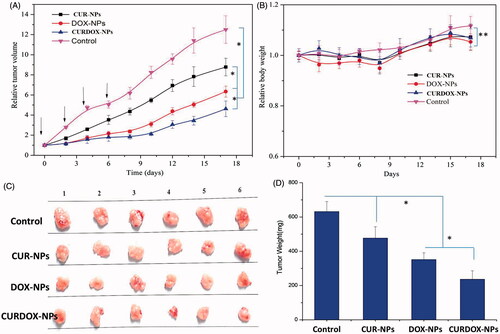
Figure 5. In vivo anti-tumour efficacy of various formulations, (A) tumour growth of MCF-7/ADR cancer xenografts treated with saline (control), CUR-NPs, DOX-NPs and CURDOX-NPs at a total drug dose of 2.5 mg/kg; (B) the loss of body weight of tumour-bearing mice after treatment; (C) tumour image and (D) tumour weight excised at 17 days post-treatment (n = 6, p < .05).