Figures & data
Figure 1. Preoperative high-resolution computed tomography (CT) of the temporal bones in case 1. (A) Axial CT view shows dislocation of the stapes superstructure from the footplate onto the promontory in the right ear. (B) Coronal view shows the inferiorly displaced stapes in the right. Arrows indicate the stapes superstructure. (C) Axial CT view for the normal left ear. Arrowhead indicates the stapes superstructure.
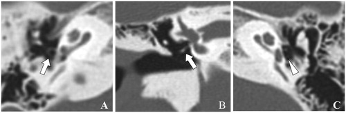
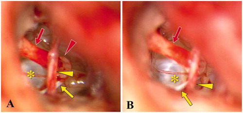
Figure 2. Intraoperative image of the middle ear in case 1. (A) The post-tympanotomy image shows the stapes superstructure attached to the promontory without formation of the stapes footplate. (B) Schema of findings in the ossicular chain and related structures. Red arrow: long process of incus, red arrowhead: stapes superstructure, yellow asterisk: oval window, yellow arrow: tendon of stapedius, and yellow arrowhead: pyramidal eminence
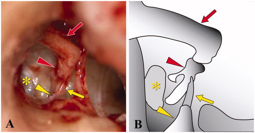
Figure 3. Preoperative high-resolution computed tomography of the temporal bones in case 2. (A, B) Axial view. Arrows indicate the inferiorly displaced stapes superstructure and arrowhead indicates the oval window portion of the utricle. (C) Coronal view shows the misattachment of the stapes superstructure on the promontory. Arrow indicates the stapes superstructure.
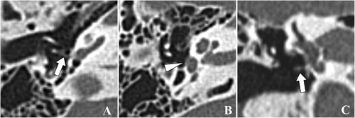
Figure 4. Schema and intraoperative microscopic view of the middle ear in case 2. (A, B) The post-tympanotomy images show the inferiorly located stapes superstructure attached to the promontory. Red arrow: long process of incus, red arrowhead: head of stapes, yellow asterisk: oval window, yellow arrow: tendon of stapedius, and yellow arrowhead: chorda tympani nerve