Figures & data
Figure 1. (A) Axial contrast enhanced (CE) computed tomographic (CT) scan, showing opacification of the inferior meatus, partly enhanced. (B) Coronal CE-CT scan. It is difficult to evaluate bone erosion or bone destruction accurately, but neither is apparent. (C) T2-weighted axial magnetic resonance imaging (MRI), showing a heterogeneous high-intensity signal in the same area as on the CT scan. (D) T1-weighted MRI with gadolinium, showing heterogeneous enhancement of the lesion.
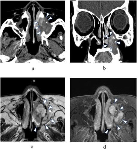
Figure 2. (A) The tumor was in the inferior meatus and was in contact with the inferior turbinate and middle turbinate. (B) Appearance after resection of the tumor. The middle turbinate (*) and inferior turbinate (**) were also resected. No characteristics of malignancy were found. (C) The resected tumor. (D) Specimen stained with hematoxylin and eosin (H&E; 40), showing sheets of atypical spindle cells. Scale bar: 50 μm. (E) Stained specimen (H&E,
200), showing several mitotic figures. Scale bar: 100 μm. (F) Immunohistochemical staining for erythroblast transformation-specific-related gene (ERG;
200). Tumor nuclei are positive for ERG, which is highly expressed in endothelial cells. Scale bar: 100 μm.
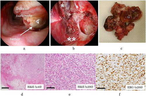
Table 1. Literature review of 23 cases of sinonasal angiosarcoma, including that of our patient.
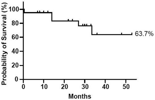
Figure 3. Kaplan–Meier survival curve representing the patients in our review, generated with GraphPad Prism 8 (GraphPad Software, San Diego, CA, USA). We calculated the probability of 3 year survival as 63.7%.