Figures & data
Figure 1. Non-contrast computed tomography demonstrates irregular growth in the thyroid lobes (curved arrow) and a nodule containing calcification foci in the right thyroid lobe (straight arrow); (a) axial, (b) sagittal, and (c) coronal planes of CT.
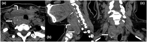
Figure 2. (a–d) FNAC of the case. Malignant endothelial cells are arranged in loosely cohesive clusters admixed with erythrocytes (E) (Papanicolau ×200).
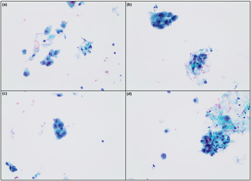
Figure 3. (a) Macroscopic image of a section from thyroid of the primary angiosarcoma case. A number of enlarged nodules are detected in different sizes, some of which are cystic formations. (b–c–d) Histopathological image of the case. Atypical cells with hyperchromatic nuclei and vascular channels containing erythrocytes are prominent (H&E, ×110, ×200, ×400).
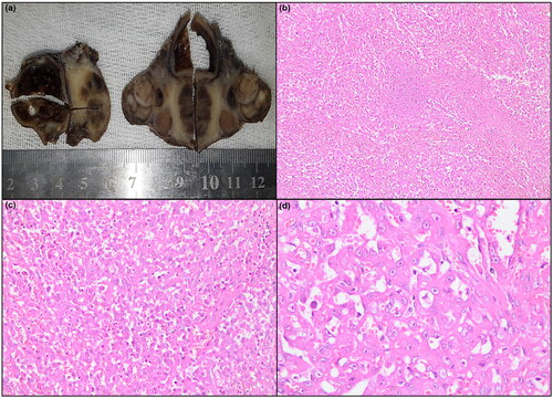
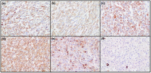
Figure 4. Immunohistochemical images of the case. (a) Positive staining of cytoplasmic granules for pan-cytokeratin (×400). (b) Positive staining of the cytoplasm for laminin (×400). (c) Positive staining of cytoplasm for vimentin (×400). (d) Positive staining of cytoplasm for CD31 (×400). (e) Positive granular staining of the cytoplasm for factor VIII (×400). (f) Negative staining for CD34 (×400).