
Figures & data
Figure 1. The setup of radiation exposure of mobile phones to the rabbits.
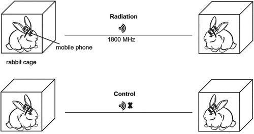
Figure 2. Effect of mobile phone radiation on the rabbit organ coefficients. Organ coefficients of control rabbits (n = 5) and radiation rabbits (n = 8).
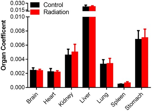
Figure 3. Histopathology analysis of rabbit tissues after 16 weeks-mobile phone radiation. Rabbit tissues were analyzed by H&E staining. Radiation, rabbits exposed to mobile phone radiation. Control, rabbits without exposure to mobile phone radiation. Normal/Lesion, normal or lesion tissues observed. Inflammatory cell infiltration (A, B, C, D) and cytoplasmic vacuolation (E) were indicated by black arrows. Representative results were shown. 200×, magnification.
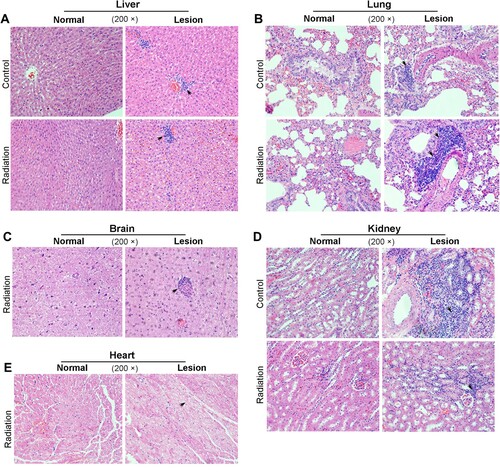
Table 1. Lesions of rabbits treated with radiation.
Figure 4. Effect of mobile phone radiation on DNA damage. Representative results of γ-H2AX staining in rabbit tissues. Radiation, exposure to mobile phone radiation. Control, sham exposure to mobile phone radiation. 200× and 400×, magnifications. (A) Brain (B) Kidney (C) Heart.
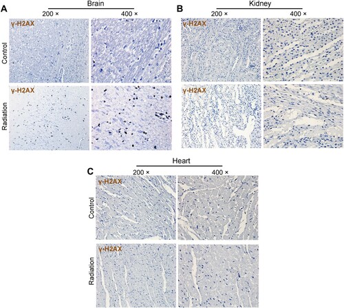
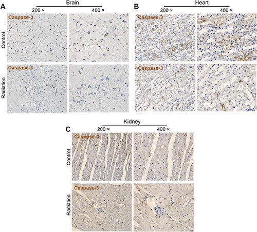
Figure 5. Effect of mobile phone radiation on cell apoptosis. Representative results of cleaved caspase-3 staining in rabbit tissues. Radiation, exposure to mobile phone radiation. Control, sham exposure to mobile phone radiation. 200× and 400×, magnifications. (A) Brain (B) Kidney (C) Heart.
Data availability statement
The data that support findings of this study are available from the corresponding author on reasonable request. The data are not publicly available at this time as the data also forms part of an ongoing study.