241
Views
2
CrossRef citations to date
0
Altmetric
Original

OXYGEN METABOLITES AND HYPOXIC RENAL INJURY: EFFECT OF MITOCHONDRIAL ELECTRON TRANSPORT BLOCKADE

, M.D. & , M.S.
Pages 249-258 | Published online: 07 Jul 2009

Figures & data

Figure 1. Electron micrographs of thick ascending limb of loop of Henle after perfusion fixation in vivo (A), following 60 min of standard perfusion (B) and after 60 min of perfusion with addition of rotenone to the standard perfusate (C). Standard perfusion causes hypoxic cell death in the tubules. Nuclei are pyknotic and cells are fragmented. These lesions are prevented by the addition of rotenone.
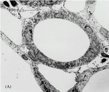
Figure 1. Electron micrographs of thick ascending limb of loop of Henle after perfusion fixation in vivo (A), following 60 min of standard perfusion (B) and after 60 min of perfusion with addition of rotenone to the standard perfusate (C). Standard perfusion causes hypoxic cell death in the tubules. Nuclei are pyknotic and cells are fragmented. These lesions are prevented by the addition of rotenone.

Figure 2. Catalase activity in the outer medulla after 10 min of isolated perfusion of the kidney under various conditions is shown. The dotted lines represent the range covered by the mean ± SEM for catalase activity in the outer medulla in vivo in untreated rats and are provided for reference. Standard kidney perfusion without aminotriazole (AMTZ) causes no statistically significant decrease in medullary tissue catalase activity from the in vivo level. The presence of AMTZ in the medium during otherwise standard perfusion results in a decrease in tissue catalase activity indicating H2O2 production during the perfusion. Addition of either rotenone or antimycin prevents the loss of catalase activity caused by the presence of AMTZ indicating inhibition of H2O2 production. ANOVA indicates p<0.001 for differences among the groups. Standard perfusions with aminotriazole (**) show statistically significant difference from all other groups by Bonferroni's method of post-test analysis. None of the other groups differ significantly from each other or from the in vivo level.
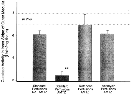
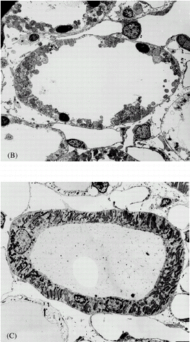
Figure 2. Catalase activity in the outer medulla after 10 min of isolated perfusion of the kidney under various conditions is shown. The dotted lines represent the range covered by the mean ± SEM for catalase activity in the outer medulla in vivo in untreated rats and are provided for reference. Standard kidney perfusion without aminotriazole (AMTZ) causes no statistically significant decrease in medullary tissue catalase activity from the in vivo level. The presence of AMTZ in the medium during otherwise standard perfusion results in a decrease in tissue catalase activity indicating H2O2 production during the perfusion. Addition of either rotenone or antimycin prevents the loss of catalase activity caused by the presence of AMTZ indicating inhibition of H2O2 production. ANOVA indicates p<0.001 for differences among the groups. Standard perfusions with aminotriazole (**) show statistically significant difference from all other groups by Bonferroni's method of post-test analysis. None of the other groups differ significantly from each other or from the in vivo level.

Reprints and Corporate Permissions

Please note: Selecting permissions does not provide access to the full text of the article, please see our help page How do I view content?

To request a reprint or corporate permissions for this article, please click on the relevant link below:

Academic Permissions

Please note: Selecting permissions does not provide access to the full text of the article, please see our help page How do I view content?

Obtain permissions instantly via Rightslink by clicking on the button below:

If you are unable to obtain permissions via Rightslink, please complete and submit this Permissions form. For more information, please visit our Permissions help page.