Figures & data
Table 1. Patient Characteristics
Table 2. Parameters of Phospholipase A2 Clearance (mean ± SD) of LP‐CVVHDF
Figure 1. Phospholipase A2 (PLA2) activity and protein concentration after heparin elution from large‐pore hemofilter. One hemofilter was washed with 1 L of acetate Ringer solution and ten 5‐mL fractions every 100 mL of washed solution (fraction number: F1 to F10) were collected. PLA2 bound to hemofilter was eluted with 200 mL of saline including 1 mg/mL heparin and two 5 mL‐fractions every 100 mL of eluting solution (fraction number: H1, H2) were collected. Protein concentration (open circle) and PLA2 activity (solid circle) of each fraction were assayed.
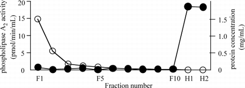
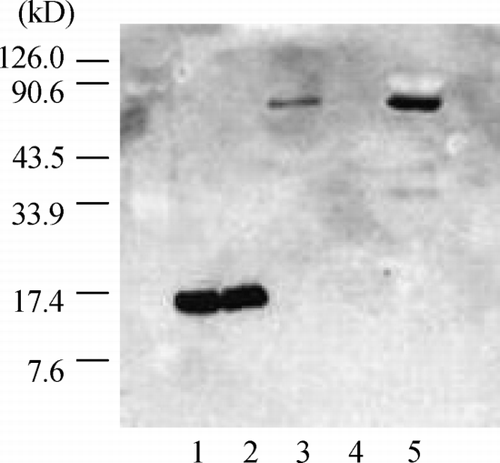
Figure 2. Western blot analysis of the eluted phospholipase A2 (PLA2) using anti‐human group IIA PLA2 monoclonal antibody. The purified IIA PLA2 (lane 1) and purified PLA2 including 1 mg/mL heparin (lane 2) were identified as one approximately 14 kD band, and the PLA2 in eluting solution (H‐1) (lane 3) was identified as an approximately 70 kD band. There was no band in washing solution (F‐10) (lane 4). The PLA2 in the ultradiafiltrate was identified as an approximately 70 kD band (lane 5).