Figures & data
Table 1. Neuroimaging instruments, types of imaging, biomarkers and biological & clinical correlates in PD.
Table 2. Comparison of neuroimaging modalities.
Figure 1. Dopamine neurons induced from autologous bone marrow mesenchymal stem cells (MSCs) in parkinsonian monkeys and post-graft PET imaging. A) Bone marrow MSCs in primate animals. B) Neuron-like cells induced from MSCs. Immunocytochemistry showed that these neuron-like cells were positive for markers of neurons, Tuj-1 (C) and MAP-2 (D), catecholamine neurons, TH (E), dopaminergic neurons, DAT (F). G) After being autologously grafted into the striatum of MPTP-treated hemi-parkinsonian animals, PET imaging showed high binding potential (BP) of the DAT ligand, 11C-CFT in the dorso-lateral part of the putamen (yellow arrow). White arrow shows high BP in the normal side of the putamen. H) Kinetic analysis of BP data obtained repeatedly till 7 months after transplantation. The kinetic analysis indicated that a major part of the grafted cells (∼ 90%) lost the expression of DAT (or degenerated) with a half-life of 10 days, while the rest of the grafted cells retained the DAT expression at the 7-month post-graft.
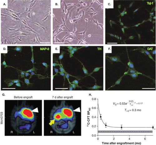
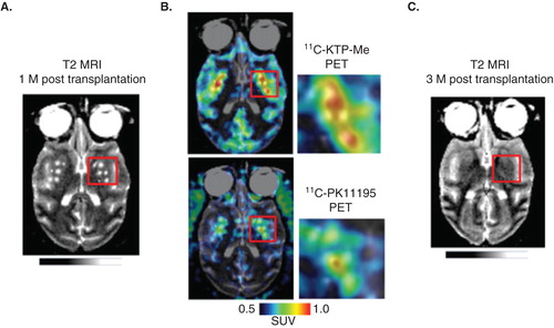
Figure 2. Potential predictability of the post-graft inflammatory response for delayed graft cell death. A. T2-weighted MRI brain image of a PD model animal, in which human embryonic stem cells were transplanted bilaterally in the striatum 1 month before MRI scan. B. At the same time point, PET ligands for activated glia, 11C-ketoprofen methyl ester (upper) and 11C-PK11195 (lower) were accumulated in the grafted areas of striatum, suggesting that host tissues have significant immune reactions to the grafted cells. C. Follow-up T2-weighted MRI scan in the same animal (3-month post-transplantation), which showed disappearance of grafts at this later stage. These findings suggest potential usefulness of multi-modal neuroimaging in predicting the long-term graft survival from early stage information of graft–host immune interaction. Unpublished data, courtesy of Prof. Jun Takahashi at Kyoto University.