Figures & data
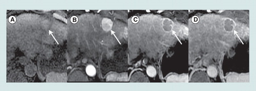
Axial T1-weighted fat-saturated magnetic resonance images obtained precontrast (A) and after injection of an extracellular gadolinium-based contrast agent, in the hepatic arterial (B), portal venous (C) and 5-min delayed (D) phases show a 2.5 cm mass in segment 2 (arrow) of the liver in a 63-year-old woman with cirrhosis due to hepatitis C viral infection. Relative to liver, the mass is mildly hyperintense precontrast, hyperenhances in the arterial phase, and hypoenhances in the portal venous and delayed phases (washout appearances). A peripheral rim of enhancement is evident in the portal venous and delayed phases (capsule appearance). In a patient with cirrhosis or other risk factors for HCC, a mass with these imaging features is categorized LI-RADS 5 (100% certainty observation is HCC).
HCC: Hepatocellular carcinoma; LI-RADS: Liver Imaging Reporting and Data System.