Figures & data
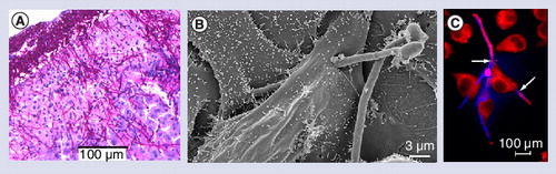
(A) Histological section showing C. albicans hyphae invading the murine liver 24 h after intraperitoneal infection. Scale bar = 100 µm. (B) Scanning electron microscopy of C. albicans invasion into oral epithelial cells after 3 h coincubation. A C. albicans hypha penetrates the epithelial cell, grows through the cell, exits the epithelial cell and invades another epithelial cell. The second hyphal cell is not invasive. Scale bar = 3 µm. Kindly provided by Gudrun Holland and Norbert Bannert (Robert Koch Institut, Berlin). (C) Fluorescence microscopy picture of human macrophages (J774) interacting with C. albicans cells after 2 h of coincubation. Red: macrophages; pink: extracellular C. albicans; blue: intracellular C. albicans. Two C. albicans hyphae have pierced a macrophage to escape (arrows). Scale bar = 100 µm.