831
Views
29
CrossRef citations to date
0
Altmetric
Review

Integrating contextual miRNA and protein signatures for diagnostic and treatment decisions in cancer

Pages 813-827 | Published online: 09 Jan 2014

Figures & data

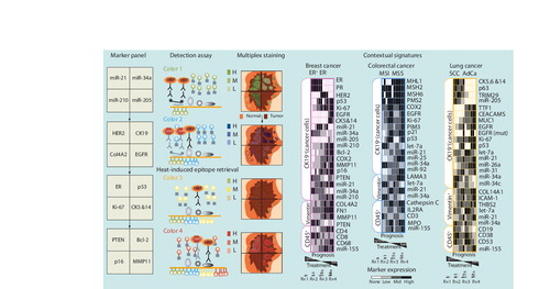
Figure 1. Contextual miRNA and protein signatures for cancer diagnostics and treatment.

On the left is a flow chart of a conceptual high-density marker detection platform. Physical compartmentalization yields four contiguous but insulated compartments for the parallel detection of independent biomarkers from the tumor core to adjacent normal tissue. Specific locked nucleid acid-modified DNA probes are dispensed in each compartment (marker panel) and tissue slides are subjected to in situ hybridization assay. miRNA signal is revealed by tyramide signal amplification (TSA) reactions with green fluorochrome-tyramine substrate (color 1; detection assay). After hydrogen peroxide incubation to inactivate HRP from the preceding TSA reaction, protein expression is revealed by a new round of TSA reactions with a different fluorochrome–tyramine substrate (color 2). After heat-induced epitope retrieval, expression of additional proteins is revealed by sequential TSA reactions with other fluorochrome–tyramine substrates (colors 3 and 4). In this example, the colorized tissue cartoon provides a virtual rendition of the expected staining pattern for each marker on an ER+PR+HER2- breast cancer specimen (multiplex staining). On the right are tentative and speculative multimarker panels based on biomarkers already incorporated in routine clinical practice and promising translational biomarkers. Computer-assisted image analysis will be used to quantitate the expression levels of each marker. This information will be used to generate contextual signatures that reflect molecular changes within the cancer cells (CK19+) or other cellular compartments of the tumor microenvironment, such as reactive fibroblast (vimentin+) and immune cells (CD45+), to inform treatment selection and intensity in prevalent solid tumors.

AdCa: Adenocarcinoma; EGFR: EGF receptor; ER: Estrogen receptor; H: High; HER2: Human EGF receptor 2; HRP: Horseradish peroxidase; L: Low; M: Medium; MMP: Matrix metalloproteinase; MSI: Microsatellite instable: MSS: Microsatellite stable; PR: Progesterone receptor; SCC: Squamous cell carcinoma.

Figure 1. Contextual miRNA and protein signatures for cancer diagnostics and treatment.On the left is a flow chart of a conceptual high-density marker detection platform. Physical compartmentalization yields four contiguous but insulated compartments for the parallel detection of independent biomarkers from the tumor core to adjacent normal tissue. Specific locked nucleid acid-modified DNA probes are dispensed in each compartment (marker panel) and tissue slides are subjected to in situ hybridization assay. miRNA signal is revealed by tyramide signal amplification (TSA) reactions with green fluorochrome-tyramine substrate (color 1; detection assay). After hydrogen peroxide incubation to inactivate HRP from the preceding TSA reaction, protein expression is revealed by a new round of TSA reactions with a different fluorochrome–tyramine substrate (color 2). After heat-induced epitope retrieval, expression of additional proteins is revealed by sequential TSA reactions with other fluorochrome–tyramine substrates (colors 3 and 4). In this example, the colorized tissue cartoon provides a virtual rendition of the expected staining pattern for each marker on an ER+PR+HER2- breast cancer specimen (multiplex staining). On the right are tentative and speculative multimarker panels based on biomarkers already incorporated in routine clinical practice and promising translational biomarkers. Computer-assisted image analysis will be used to quantitate the expression levels of each marker. This information will be used to generate contextual signatures that reflect molecular changes within the cancer cells (CK19+) or other cellular compartments of the tumor microenvironment, such as reactive fibroblast (vimentin+) and immune cells (CD45+), to inform treatment selection and intensity in prevalent solid tumors.AdCa: Adenocarcinoma; EGFR: EGF receptor; ER: Estrogen receptor; H: High; HER2: Human EGF receptor 2; HRP: Horseradish peroxidase; L: Low; M: Medium; MMP: Matrix metalloproteinase; MSI: Microsatellite instable: MSS: Microsatellite stable; PR: Progesterone receptor; SCC: Squamous cell carcinoma.

Table 1. Diagnostic and prognostic applications of miRNA signatures in solid tumors.

Table 2. Diagnostic and prognostic applications of single miRNAs in solid tumors.
Reprints and Corporate Permissions

Please note: Selecting permissions does not provide access to the full text of the article, please see our help page How do I view content?

To request a reprint or corporate permissions for this article, please click on the relevant link below:

Academic Permissions

Please note: Selecting permissions does not provide access to the full text of the article, please see our help page How do I view content?

Obtain permissions instantly via Rightslink by clicking on the button below:

If you are unable to obtain permissions via Rightslink, please complete and submit this Permissions form. For more information, please visit our Permissions help page.