
Figures & data
Table 1 Classification of Rectovaginal Fistulas
Figure 1 The schematic diagram of the female pelvis (sagittal section) showing the rectovaginal septum (yellow dotted line), type-I rectovaginal fistula (involving rectovaginal septum) (blue color), and type-II rectovaginal fistula (not involving rectovaginal septum) (red color).
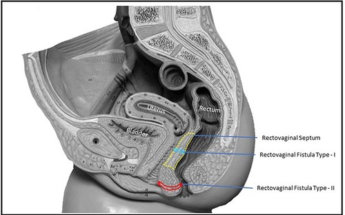
Table 2 Garg Incontinence Scoring (GIS) system
Figure 2 The graph of preoperative and postoperative anal manometry in a 25-year-old female patient who underwent fistulotomy for rectovaginal fistula with no rectovaginal septum involvement (RVF-II) fistula.
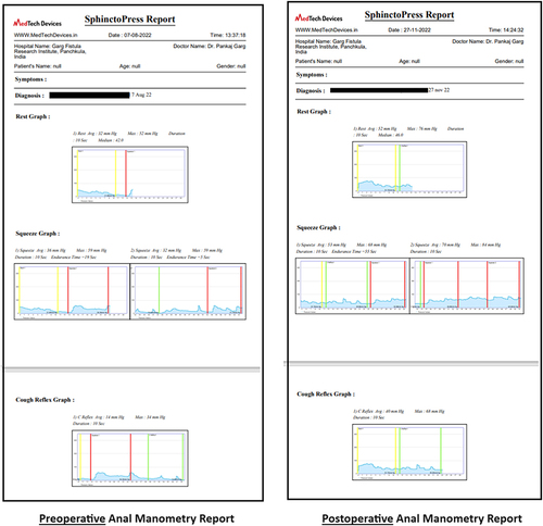
Table 3 Demography and Fistula Characteristics
Figure 3 A 33-year old female patient with anterior horseshoe, anterior anal fistula and a RVF-II fistula. She underwent a fistulotomy. Upper panel: Schematic diagram of coronal (left side) and axial sections (right side). Middle panel: Preoperative magnetic resonance imaging - Axial sections: T2 weighted (left side) and Short Tau Inversion Recovery (STIR) (right side) (yellow arrows showing fistula tract). Lower panel: Postoperative magnetic resonance imaging- 3 months after surgery - Axial sections: T2 weighted (left side) and Short Tau Inversion Recovery (STIR) (right side showing the complete healing of the fistula.
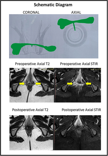
Figure 4 A 36-year old female patient with posterior horseshoe anal fistula and an RVF-II fistula opening in the left posterior vagina. She underwent the TROPIS procedure. Upper panel: Schematic diagram of coronal (left side) and axial sections (right side). Middle panel: Preoperative magnetic resonance imaging - Axial sections: T2 weighted (left side) and Short Tau Inversion Recovery (STIR) (right side) (yellow arrows showing fistula tract, green arrows showing fistula tract opening into vagina). Lower panel: Postoperative magnetic resonance imaging- 2 months after surgery - Axial sections: T2 weighted (left side) and Short Tau Inversion Recovery (STIR) (right side showing the complete healing of the fistula.
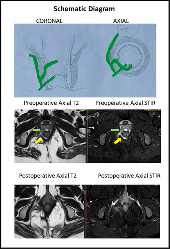
Table 4 Change in Objective Incontinence Scores (Garg Incontinence Scores) After Surgery
Table 5 Change in Anal Manometry After Surgery