Figures & data
Figure 1 Theories of posterior semicircular canal stimulation in BPPV.
Notes: (A) Cupulolithiasis: adherence of debris to the cupula making it gravity sensitive (1. Sensory epithelium. 2. Cupula. 3. Otoconia. 4. Ampulla). (B) Cupulolithiasis: cupula deflection after canal rotation. (C) Canalolithiasis: position of particles before canal rotation. (D) Canalolithiasis: movement of particles and cupula deflection after canal rotation.
Abbreviation: BPPV, benign paroxysmal positional vertigo.
Abbreviation: BPPV, benign paroxysmal positional vertigo.
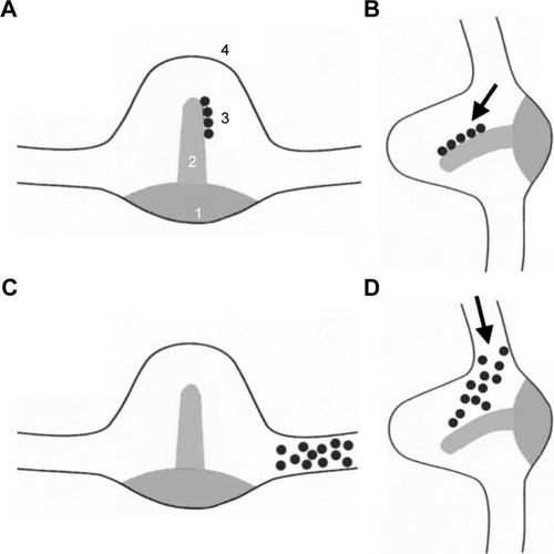
Figure 2 The Dix-Hallpike test, for the left (L) ear on the left panel and for the right (R) ear on the right panel.
Notes: (A) Initial sitting position. (B) Head turn 45° toward the examined ear. (C) Head hanging 45° below the horizontal, with the examined ear undermost.
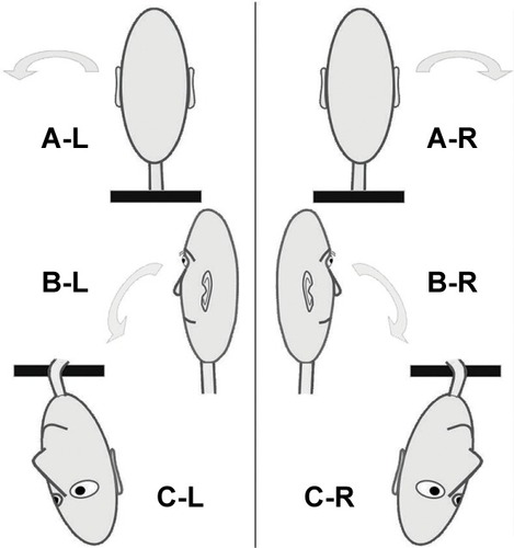
Table 1 Diagnosis of the involved semicircular canal and the side of involvement, according to the appropriate diagnostic maneuver
Table 2 Diagnostic test, characteristics and treatment of BPPV types
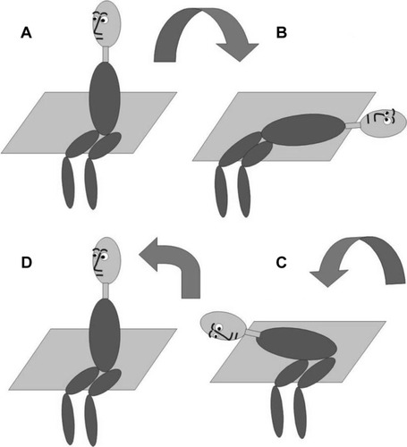
Figure 3 The Epley canalith repositioning procedure when the posterior semicircular canal of the left ear is affected.
Notes: (A) The patient is sitting with the head turned horizontally 45° to the affected (left) ear. (B) Left head-hanging position. (C) Rightward roll, right head-hanging position. (D) Further rightward roll. (E) Sitting up. The head is held still in all positions for 1–3 minutes.
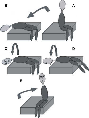
Figure 4 The Semont maneuver when the posterior semicircular canal of the left ear is affected.
Notes: (A) The patient sits with the head turned horizontally 45° to the healthy (right) ear. (B) Moving to left side-lying position (nose up). (C) Moving to the right side-lying position (nose down). Then the patient returns to initial position (D). Again the head is held still in all positions for 1–3 minutes.