Figures & data
Figure 1 Assay of Aβ40/42 and amyloid precursor protein.
Notes: (A) The levels of Aβ40/42 proteins were measured by ELISA according to the manufacturer’s instruction. The expression of Aβ40/42 protein in the minocycline treatment group was significantly decreased when compared with the control model group. *Denotes P < 0.01 versus control group. (B) The levels of amyloid precursor protein were measured by immunostaining (1:200, Biosource International, Inc, Camarillo, CA, USA). It seems that there is no significant difference between the control model group and the minocycline intervention group. *Denotes P > 0.05 versus control group. The model group was subdivided into M4, M6, and M8 for 4, 6, and 8 weeks after streptozocin injection, and the minocycline administration group into MT4, MT6, and MT8.
Abbreviations: APP, amyloid precursor protein; M4, M6, M8: 4, 6, and 8 weeks after establishment of diabetes; MT4, MT6, MT8: 4, 6, and 8 weeks after minocycline intervention; OD, optical density.
Abbreviations: APP, amyloid precursor protein; M4, M6, M8: 4, 6, and 8 weeks after establishment of diabetes; MT4, MT6, MT8: 4, 6, and 8 weeks after minocycline intervention; OD, optical density.
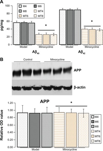
Figure 2 Assay for tau proteins.
Notes: (A) The levels of total tau protein (Bioss Co, Beijing, People’s Republic of China) by western blotting (1:200) or immunohistochemistry (1:100) showed no significant change between the control and minocycline intervention group. *Denotes P > 0.05 versus control group. (B) The levels of phosphorylated tau proteins by western blotting as expressed by the densitometry ratio of tau proteins to β-actin (mean ± standard deviation) were measured, including pre-tangle marker phospho-tau antibody TG3 (pT231), intraneuronal tangle marker phospho-tau protein (Ser214, pS214), and extracellular tangle marker PHD finger protein-1 (pS396/pS404). These results showed that the optical density value of phospho-tau proteins significantly decreased after minocycline treatment (P = 0.0001). pT231 (1:200), ps214 (1:200), ps396 (1:100), and ps404 (1:100) were purchased from Bioss Co, scale bar is 20 μm.
Abbreviations: OD, optical density; M4, M6, M8: 4, 6, and 8 weeks after establishment of diabetes; MT4, MT6, MT8: 4, 6, and 8 weeks after minocycline intervention; PHF-1, PHD finger protein-1.
Abbreviations: OD, optical density; M4, M6, M8: 4, 6, and 8 weeks after establishment of diabetes; MT4, MT6, MT8: 4, 6, and 8 weeks after minocycline intervention; PHF-1, PHD finger protein-1.
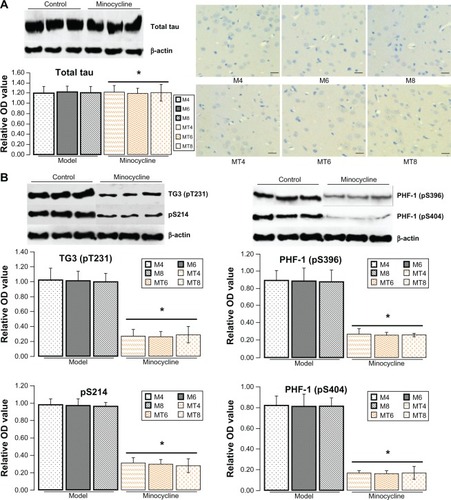
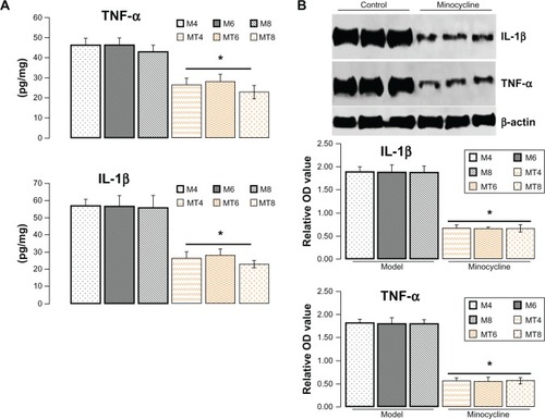
Figure 3 Assay of protein level of interleukin-1β and tumor necrosis factor-α.
Notes: (A) The levels of interleukin (IL)-1β and tumor necrosis factor (TNF)-α by enzyme-linked immunosorbent assay in the minocycline treatment group decreased more significantly than in the control model group. *Denotes P < 0.001. (B) The levels of IL-1β and TNF-α by western blotting, as expressed by the densitometry ratio of IL-1β and TNF-α to β-actin (mean ± standard deviation), was more evidently reduced in the minocycline treatment group than in the control model group. *Denotes P < 0.01 versus control model group.
Abbreviations: IL-1β, interleukin-1β; OD, optical density; TNF-α, tumor necrosis factor-α; M4, M6, M8: 4, 6, and 8 weeks after establishment of diabetes; MT4, MT6, MT8: 4, 6, and 8 weeks after minocycline intervention.
Abbreviations: IL-1β, interleukin-1β; OD, optical density; TNF-α, tumor necrosis factor-α; M4, M6, M8: 4, 6, and 8 weeks after establishment of diabetes; MT4, MT6, MT8: 4, 6, and 8 weeks after minocycline intervention.