
Figures & data
Figure 1 An example of target lobe selection on the basis of dual-source computed tomography.
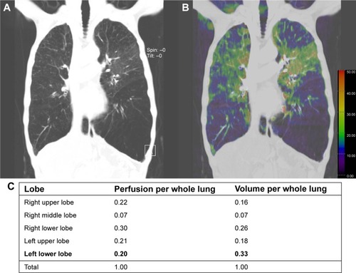
Table 1 Baseline characteristics of the patients
Figure 2 Flow chart indicating patient disposition in the study.
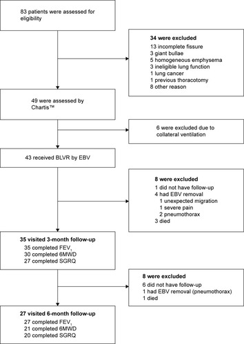
Figure 3 Primary outcomes 3 and 6 months after endobronchial valve therapy relative to baseline.
Abbreviations: FEV1, forced expiratory volume in 1 second; 6MWD, 6-minute walk distance test; mMrc, modified Medical Research Council dyspnea scale; SGRQ, St George’s Respiratory Questionnaire.
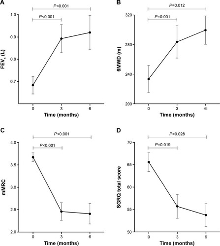
Figure 4 Proportion of patients whose clinical outcomes after endobronchial valve therapy exceed the minimum clinically important differences FEV1, 6MWD and SGRQ.
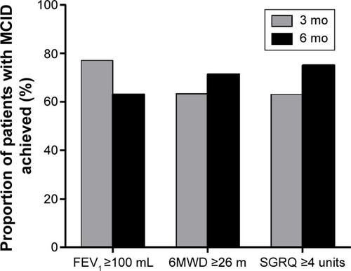
Table 2 Primary outcomes relative to baseline in patients who did and did not achieve target lobe volume reduction
Table 3 Adverse events in the 6 months after bronchoscopic lung volume reduction with endobronchial valve. (n=43)
Table 4 Comparison of our study results with those of previous trials in Western countries in terms of pneumothorax frequency and baseline characteristics
Table S1 Pulmonary function test, 6MWD, mMRC dyspnea scale, and SGRQ total score results 3 and 6 months after endobronchial valve therapy
Table S2 Effect of tuberculosis scar on target lobe volume reduction and pneumothorax