
Figures & data
Figure 1 Search strategy flow diagram for PVR and steroids.
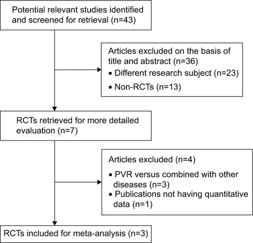
Table 1 Patient characteristics in the included trials
Figure 2 Methodological quality summary. Authors’ judgments about each methodological quality item for each included study.
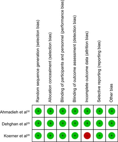
Figure 3 Forest plot of the postoperative PVR incidence rate.
Abbreviations: CI, confidence interval; M–H, Mantel–Haenszel test; PVR, proliferative vitreoretinopathy.
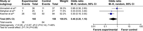
Figure 4 Forest plot of the postoperative PVR incidence rate (excluding grade C, Ahmadieh et al study). There was a statistically significant difference between the steroid group and the control group, when the study of PVR grade C was excluded.
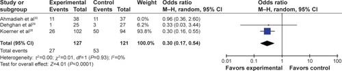
Figure 5 Forest plot of visual acuity evaluated at 6 months post-RRD surgery. There was no difference in visual acuity between the steroid group and the control group.
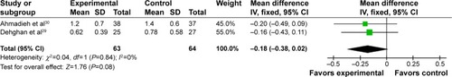
Table 2 Outcome indicators in the included trials