Figures & data
Table 1 Mean IOP changes
Table 2 MDA, NO, and NOS2 levels in the study groups
Figure 1 Micrographs of retinal TUNEL staining for one rat in each treatment group.
Notes: (A) Control, (B) vehicle control, and (C) ghrelin. Arrows indicate apoptotic cells.
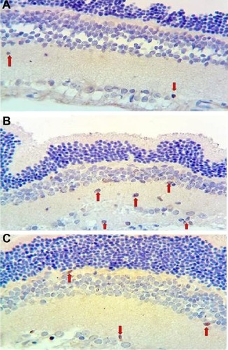
Figure 2 Micrographs of hematoxylin-eosin staining for one rat in each treatment group.
Notes: (A) Control, (B) vehicle control, and (C) ghrelin. Arrows indicate the INL.
Abbreviations: ONL, outer nuclear layer; OPL, outer plexiform layer; INL, inner nuclear layer; IPL, inner plexiform layer; RGC, retinal ganglion cell.
Abbreviations: ONL, outer nuclear layer; OPL, outer plexiform layer; INL, inner nuclear layer; IPL, inner plexiform layer; RGC, retinal ganglion cell.
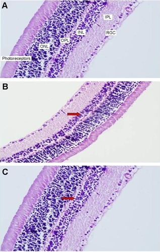
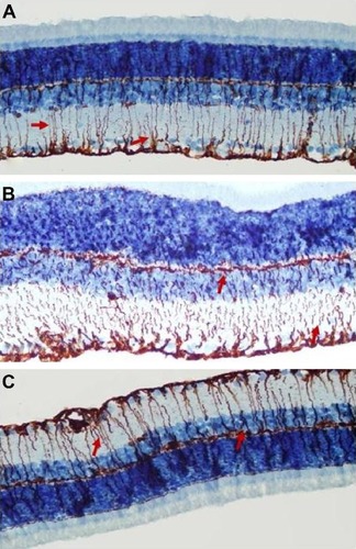
Figure 3 Micrographs of retinal glial fibrillary acid staining for one rat in each treatment group.
Notes: (A) Control, (B) vehicle control, and (C) ghrelin. Arrows indicate Müller cells and their processes.
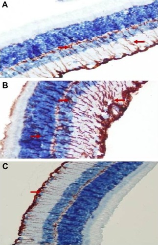
Figure 4 Micrographs of retinal S-100 staining for one rat in each treatment group.
Notes: (A) Control, (B) vehicle control, and (C) ghrelin. Arrows indicate Müller cells and their processes.
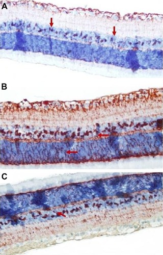
Figure 5 Micrographs of retinal vimentin staining for one rat in each treatment group.
Notes: (A) Control, (B) vehicle control, and (C) ghrelin. The arrows indicate Müller cells and their processes.