Figures & data
Table 1 Different Groups of Cell Treatment
Figure 1 C2C12 cells on the third day of differentiation, inverted microscopy (A) ×100; (B) ×200.
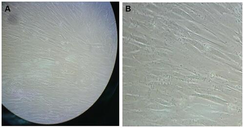
Figure 2 (A) GLUT-4 bands of membrane fractions, (B) GLUT-4 bands of cytosolic fractions. Control: cells treated with 50 μg/mL DMSO, but without any inhibitor. Treatment 1: cells treated with 50 μg/mL ginger extract without any inhibitor. Treatment 2: cells treated with 50 μg/mL ginger extract and 20 μM AMPK inhibitor. Treatment 3: cells treated with 50 μg/mL ginger extract and 25 μM PI3K inhibitor.
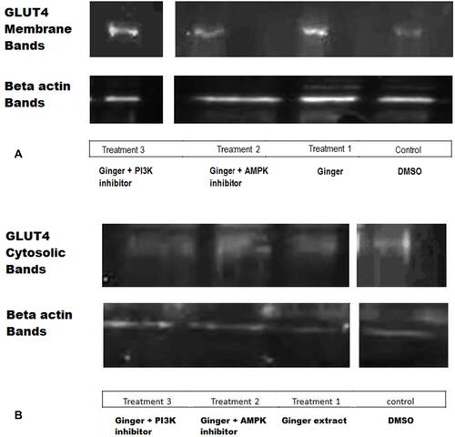
Figure 3 Comparison of GLUT-4 quantity between cytosolic and membrane fractions. In each treatment group there was significantly more amount of GLUT-4 protein in membrane fractions in comparison to cytosolic fractions, P value = 0.012 by Bonferroni post hoc test.
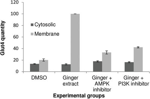
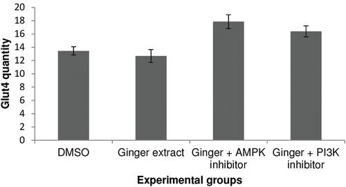
Figure 4 Comparison of GLUT-4 quantity between membrane fractions of different treatment groups. The amount of GLUT-4 protein was significantly more in membrane fraction of treatment 1 (cells treated with 50 μg/mL ginger extract without any inhibitors) compared to the control (cells treated with 50 μg/mL DMSO without any inhibitors) (p = 0.013), and also higher in comparison with the treatment 2 (cells treated with 50 μg/mL ginger extract and 20 μM compound C) and treatment 3 (cells treated with 50 μg/mL ginger extract and 25 μM LY294002) groups.
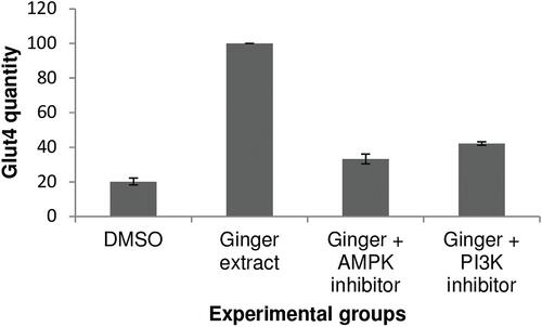
Figure 5 Comparison of GLUT-4 quantity between cytosolic fractions of different treatment groups. There was significantly more amount of GLUT-4 protein in cytosolic fraction of treatment 2 (cells treated with 50 μg/mL ginger extract and 20 μM compound C) in comparison with the treatment 1, P value = 0.039. A similar situation was seen in cytosolic fraction of treatment 3 (cells treated with 50 μg/mL ginger extract and 25 μM LY294002) compared to the treatment 1, but there was no significant different between treatment 2 and treatment 3, and also between control and treatment 1 (P value > 0.05).