Figures & data
Figure 1 Outline scheme of the experimental design used in this study.
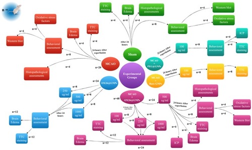
Figure 2 Physiochemical characterization of GNPs. (A) Particle size analysis of OX26@GNPs using dynamic light scattering (DLS), (B) Zeta potential distribution of GNPs along with OX26@GNPs. TEM images of (C) GNPs (scale bar 200 nm) and (D) OX26@GNPs (scale bar 100 nm).
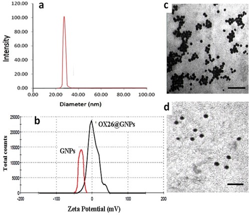
Table 1 Hydrodynamic Size (nm) Of Bare And OX26@GNPs In Different Aqueous Media
Table 2 The Zeta Potential (mv) Of Bare And OX26@GNPs In Different Aqueous Media
Figure 3 The Au content in the different tissues of male Wistar rats at 24 h after intravenous injection of 500 μg/mL PEGylated GNPs and OX26@GNPs (**P<0.01 between indicated groups).
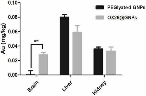
Figure 4 The effect of OX26@GNPs on infarct volume and neurological deficits. a) Representative images of infarct size at 72 h post-MCAO by TTC staining in the sham, MCAO and OX26@GNPs+MCAO (500 μg/mL, i.v. dose of GNPs) (**P<0.01 between indicated groups, n=5, #P<0.05 between indicated groups). b) neurological deficit score at 72 h post-MCAO (#P<0.05 between indicated groups), and c) ladder test (*P<0.05, #P<0.05, ###P<0.001 between indicated groups, data presented as means ± SEMs).
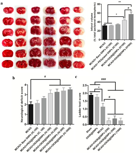
Figure 5 Nissl-stained brain sections of the CA1subregion of the hippocampus 72 h post-MCAO at a 500 μg/mL dose of OX26@GNPs (magnification 200×). *: macro picture demonstrates the region where the assessments were done. (A) Sham, (B) MCAO, and (C) MCAO+ OX26@GNPs. The green arrows demonstrate intact cells and red arrows demonstrate necrotic cells. The graph demonstrates the density of intact neuronal cells in the hippocampal CA1subregion (***P<0.001, ##P<0.01, ###P<0.001 between indicated groups).
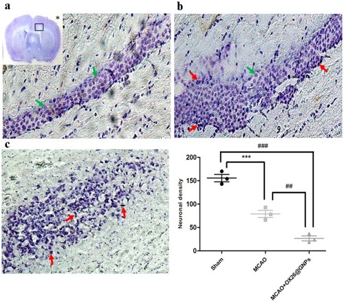
Figure 6 The effects of OX26@GNPs on: (A) Brain edema (*P<0.05, #P<0.05, ###P<0.001 between indicated groups), (B) brain LDH activity (**P<0.01, #P<0.05, ###P<0.001 between indicated groups), (C) food intake ($$P<0.01 in 1 and 2 days, $ P<0.05 in 3–6 days vs. sham and OX26@GNPs groups; ###P<0.001 in 1–14 days vs. sham and OX26@GNPs groups; *P<0.05 in 13 and 14 days vs. MCAO+OX26@GNPs group, 500 μg/mL, i.v.), D) body weight changes (# P<0.05 in 3–1 days; *P<0.05 and ## P<0.01 in 7–1 days between indicated groups).
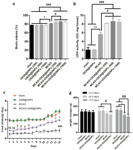
Figure 7 The effects of OX26@GNPs administration (500 μg/mL, i.v.) on: (A) WBC count, (B) RBC count, (C) AST, (D) ALT, (E) urea, and (F) creatinine. Data presented as mean ± SD.
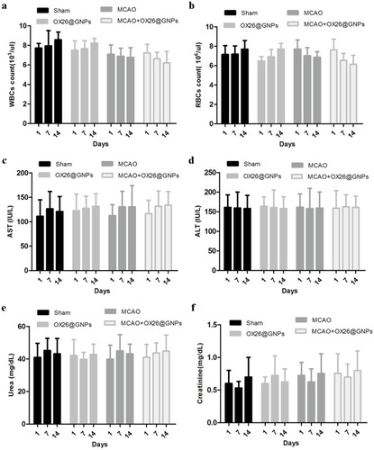
Figure 8 The effects of OX26@GNPs (500 μg/mL, i.v.) on the (A) heart, (B) lung, (C) spleen, (D) liver, (E) kidney and (F) testis 7 days post-injection (magnification 200×). The effects of OX26@GNPs (500 μg/mL, i.v.) 24 h post-MCAO in experimental groups on the expression levels of (G) RIPK1, (H) RIPK3, and (I) MLKL (*P<0.05, ##P<0.01, # P<0.05, and ###P<0.001 between indicated groups). Data presented as mean ±SEM.
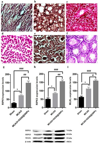
Figure 9 The effects of OX26@GNPs (500 μg/mL, i.v.) 24 h post-MCAO in experimental groups on: (A) GSH content, (B) GPx, (C) SOD, (D) CAT and (E) MDA in brain tissue under oxidative stress (***P<0.001, **P<0.01, *P<0.05, #P<0.05, ###P<0.001 and ## P<0.01 between indicated groups).
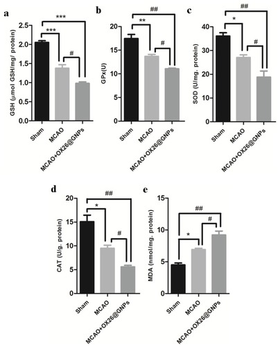
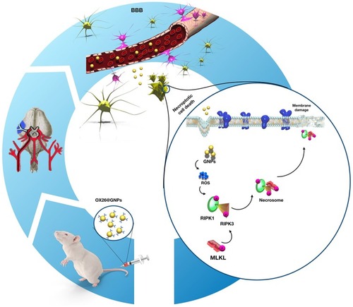
Figure 10 Proposed signaling pathways involved in OX26@GNPs-induced toxicity in a rat model of MCAO.