Figures & data
Figure 1 Physical characterization of chitosan nanoparticles. Zeta potential of (A) Sham chitosan nanoparticles and (B) Methylglyoxal-conjugated chitosan nanoparticles (MGCN). Fourier Transform Infrared Spectra of (C) Sham chitosan nanoparticles and (D) Arrow indicates the formation of imine bond in MGCN (Nano-MG).
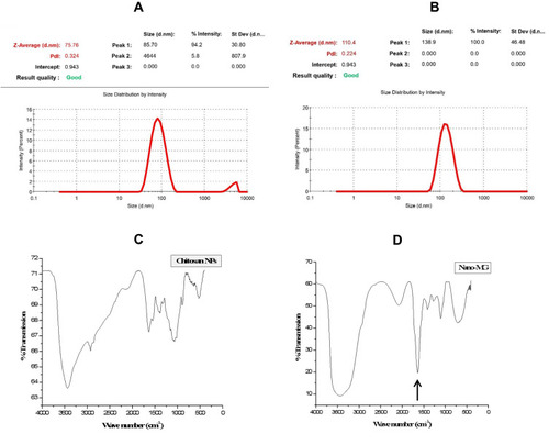
Figure 2 Transmission electron microscopy (TEM) images of (A) Sham chitosan nanoparticles and (B) Methylglyoxal-conjugated chitosan nanoparticles (MGCN). Scanning electron microscopy (SEM) images of (C) Sham Chitosan nanoparticles and (D) Methylglyoxal-conjugated chitosan nanoparticles (MGCN).
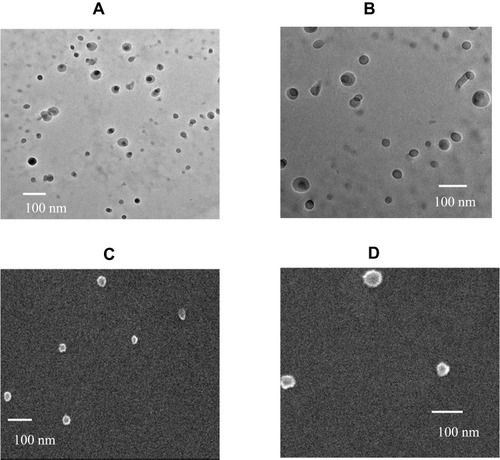
Figure 3 Anti-Candida activity of MG by agar well diffusion method. (A) Vehicle control, (B) 12.5 µg, (C) 25 µg and (D) 50 µg of MG.
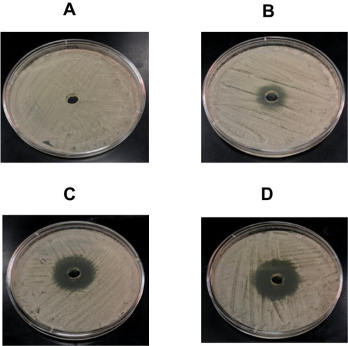
Figure 4 Effect of Fluconazole, MG and MGCN on the transition of Candida albicans yeast cells into hyphae form.
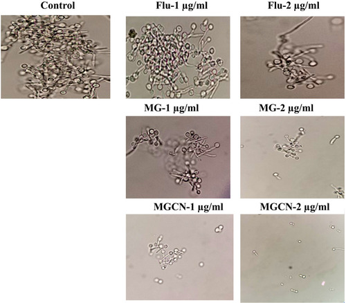
Figure 5 Macrophages (1 × 10⁶ cells/well) were infected with C. albicans at multiplicity of infection of 2 (MOI=2) followed by treatment with fluconazole or free MG or MGCN at the concentrations of 10 and 20 µM. After 48 h, the cells were lysed and intracellular yeasts were plated on Saboraud dextrose agar plates to determine the number of CFUs. Data are expressed as mean ± SE. A P value <0.05 was considered to be significant.
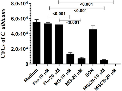
Figure 6 Fluconazole did not show antifungal activity against C. albicans. (A) Each mouse was infected with 7 x 10⁵ CFUs of C. albicans. Mice were treated with 5, 10, 20 mg/kg of fluconazole on days 1, 3 and 5 postinfection. Mice were observed for 40 days to monitor their survival. Saline (●), fluconazole (5 mg/kg) (○), fluconazole (10 mg/kg) (♦), fluconazole (20 mg/kg) (♦). (B) On day 4, three mice from each group were sacrificed and their kidney was taken to prepare tissue homogeninate. The kidney tissue homogenate was cultured in order to determine the fungal load. Data are expressed as mean ± SE. A P value <0.05 was considered to be significant.
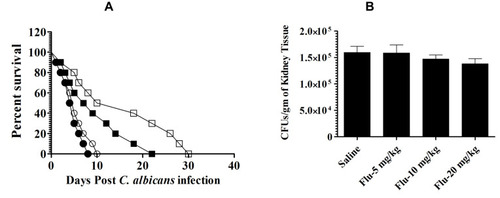
Figure 7 Treatment with MGCN showed the highest antifungal activity against C. albicans. (A) Mice infected with C. albicans (7 × 10⁵ CFUs/mouse) were treated with 1, 5 and 10 mg/kg of MG or MGCN on days 1, 3 and 5 postinfection. Mice were observed for 40 days to monitor their survival. Saline (○), SCN (●), Free MG-1 mg/kg (∆), MGCN-1 mg/kg (▲), Free MG-5 mg/kg (♦), MGCN-5 mg/kg (♦), Free MG-10 mg/kg (□), MGCN-10 mg/kg (■). Saline vs SCN (P=0.0064), Free MG-1 mg/kg vs MGCN-1 mg/kg (P=0.0284), Free MG-5 mg/kg vs MGCN-5 mg/kg (P=0.0298), Free MG-10 mg/kg vs MGCN-10 mg/kg (P=0.0258). (B) On day 4, three mice from each group were sacrificed and their kidney was taken to make tissue homogenate. The kidney tissue homogenate was cultured to determine the fungal load. A P value <0.05 was considered to be significant. *** (P<0.001).
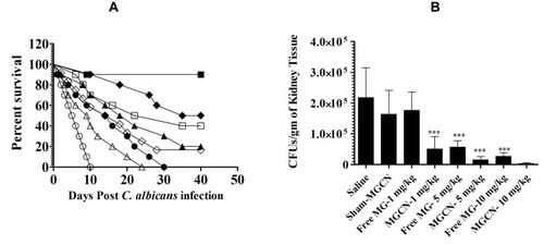
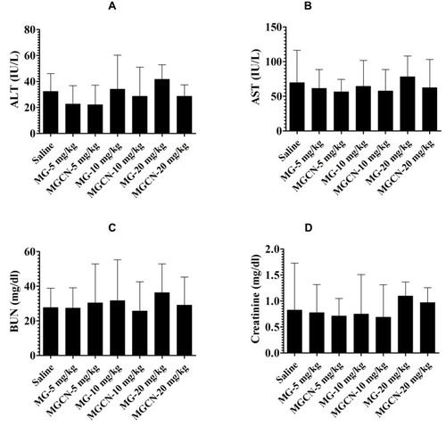
Figure 8 Administration of free MG or MGCN did not induce any toxicity. Mice were injected with MG or MGCN at the doses of 5, 10 and 20 mg/kg for consecutive 5 days. On day 6 postinjection, the blood was taken from three mice of each group to estimate the levels of (A) ALT, (B) AST, (C) BUN and (D) creatinine. Data are expressed as mean ± SE.