Figures & data
Table 1 Composition and Characteristics of Different Formulations
Figure 1 TEM images of the sertaconazole-loaded vesicles displaying (A) liposomes, (B) mucoadhesive liposomes.
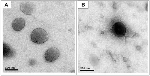
Figure 2 Entrapment efficiency of different formulae (n=3). ****P < 0.0001.
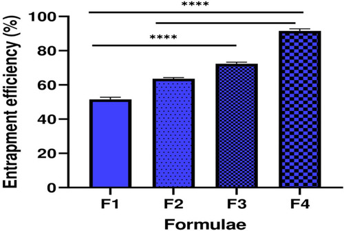
Figure 3 Particle size diameter of different formulae (n=3). *P < 0.05, **P < 0.01, ***P < 0.001, and ****P < 0.0001.
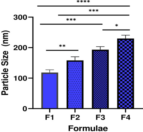
Figure 4 Zeta potential shift of mucin upon mixing with different mucoadhesive liposomes.
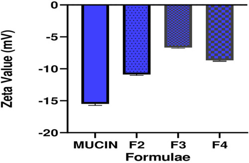
Figure 5 In vitro drug release profile.
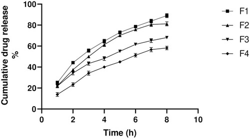
Table 2 Ex vivo Permeation Parameters of Mucoadhesive Liposomal Gel and Control Gel
Figure 6 Ex vivo permeation drug profile.
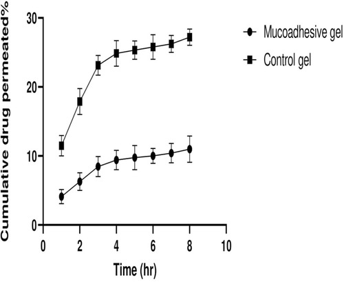
Figure 7 Cumulative drug release and tissue retention of mucoadhesive liposomal gel and control gel.
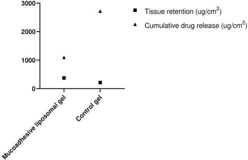
Figure 8 Mean colony-forming unit (CFU) of each group before and after the treatment period (n=6). ***P < 0.001.
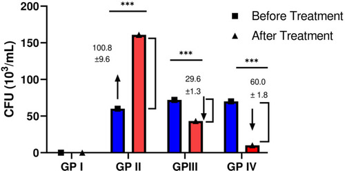
Figure 9 Assessment of serum Interleukin-23 (IL-23), beta-D-glucan (BDG), tumor necrosis factor-alpha (TNF-α), Immunoglobulin G (IgG) and Immunoglobulin M (IgM) (n=6). *P < 0.05, **P < 0.01, ***P < 0.001, and ****P < 0.0001.
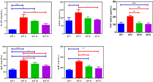
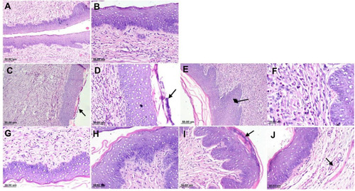
Figure 10 Photomicrograph of rat vagina, H&E stain: (A) Negative control group, normal histology of rat vagina; (B) negative control group, higher magnification, showing stratified squamous epithelium with dense sub-epithelial connective tissue; (C) positive control group, showing heavy subepithelial inflammatory cells infiltration with necrotic debris over the mucosal surface (arrow); (D) positive control group, higher magnification, showing dissolution of keratin layer with presence of necrotic tissue debris; (E) positive control group, showing hyperplastic mucosa (arrow); (F) positive control group, showing sub-epithelial neutrophils and mononuclear infiltration; (G) mucoadhesive liposomal gel group, showing mild sub-epithelial inflammatory cells infiltration; (H) showing intact mucosa; (I) sertaconazole control gel, showing mucosa with presence of necrotic debris in the keratin layer and (J) showing mild sub-epithelial edema with dilated blood vessels (arrow).