Figures & data
Table 1 Physicochemical properties of nanoparticles
Figure 1 Transmission electron microscopy images of diflucortolone valerate–loaded nanoparticles at magnification ratio of 33,000× (A), at magnification ratio of 71,000× (B).
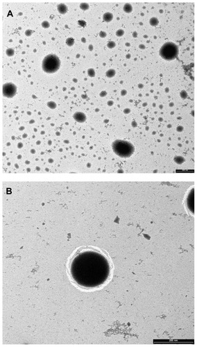
Figure 2 Differential scanning calorimetry thermograms of pure DFV (A), blank nanoparticles (B), physical mixture of blank nanoparticles and DFV (C), and drug-loaded nanoparticles (D).
Abbreviation: DFV, diflucortolone valerate.
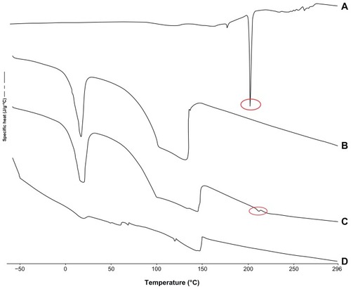
Figure 3 The in-vitro drug-release profile of diflucortolone valerate from nanoparticles (n = 6).
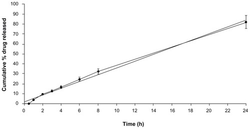
Table 2 Stability results of the nanoparticle formulations
Figure 4 Flow rheograms of chitosan gel formulations – shear rate versus shear stress (n = 3).
Abbreviations: DFV, diflucortolone valerate; Pa, pascal.
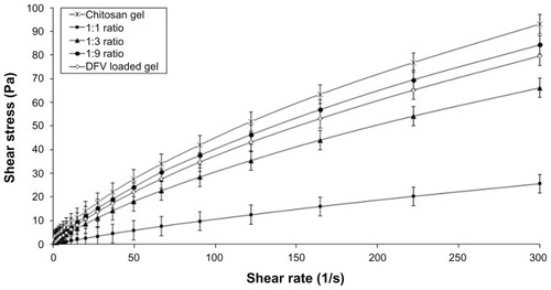
Figure 5 The elastic modulus plotted versus frequency of formulations (n = 3).
Abbreviation: DFV, diflucortolone valerate; G′, elastic (storage) modulus.
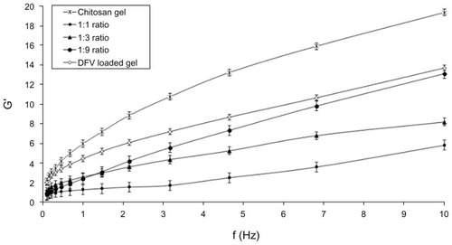
Table 3 Mechanical properties of formulations
Figure 6 Amount of DFV accumulated from formulations in the SC+epidermis (dark bars) and dermis (light bars) (n = 6).
Note: *Denotes significantly different from commercial cream and chitosan gel (P < 0.05).
Abbreviations: DFV, diflucortolone valerate; SC, stratum corneum.
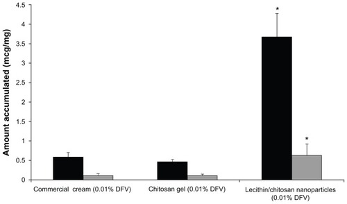
Figure 7 Accumulation of DFV from nanoparticles in chitosan gels as a function of drug concentration in the SC+epidermis (dark bars) and dermis (light bars) (n = 6).
Abbreviations: DFV, diflucortolone valerate; SC, stratum corneum.
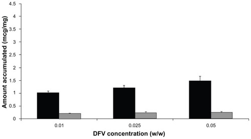
Figure 8 Changes in the paw thickness of the rats with respect to time after formulation application (n = 6).
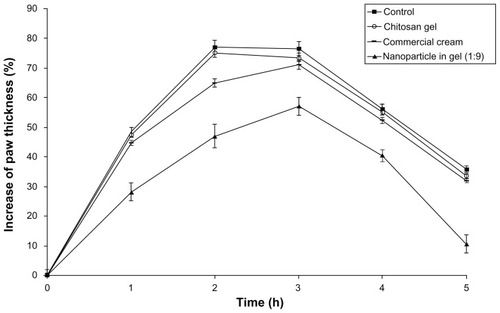
Figure 9 Skin blanching profiles of formulations – the value of color difference (Δa) in rats versus time (n = 6).
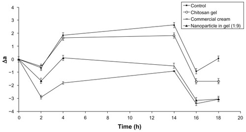
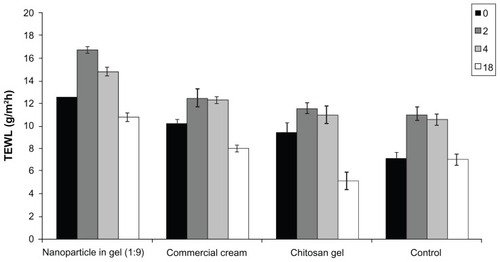
Figure 10 Changes in TEWL values of the rats with respect to time after formulation application (n = 6).
Abbreviation: TEWL, transepidermal water loss.