Figures & data
Figure 1 Overall mortality of Drosophila embryos after microtransfer of nanomaterials.
Notes: Overall mortality (OM) is the sum of the three mortality-scoring criteria. OM values are normalized against the OM of the corresponding diluent (nanomaterial OM–diluent OM). Treatments with IO nanoparticles are normalized against H2O, and Ag, Au, TiO2, SWCNT, and MWCNT are normalized against BSA 10%. For each condition tested, n=50. *Statistical relevance was established through Fisher’s exact test (P<0.005).
Abbreviations: IO, iron oxide; SWCNT, single-wall carbon nanotube; MWCNT, multiwall carbon nanotube; BSA, bovine serum albumin; Ag, silver; Au, gold; TiO2, titanium dioxide; H2O, water; Cop, coprecipitation; Thermo, thermal decomposition.
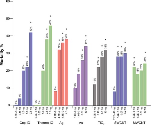
Table 1 Nanomaterial delivery amounts and dosage quantifications
Figure 2 Drosophila life cycle.
Notes: All stages of the Drosophila life cycle are readily accessible and amenable to manipulation with a variety of basic to high-end tools and techniques. Imaging techniques can be applied in every stage of development (A–H), thanks to the clear cuticle during embryonic (B and C) and larval (D–F) stages. Here we use stage 15 embryos (C), which correspond to roughly 50% of completed embryonic development. Under ideal growing conditions, this stage is reached approximately 12 hours after egg laying and features a developing central nervous system (orange), digestive tract (green and red), and many other systems (not shown) with development underway (I). In stage 15, the midgut has one compartment that divides into two distinct compartments as the embryo progresses to stage 16. We used this feature as an indication of initial survival after nanoparticle delivery and used morphological features characteristic of later developmental stages (J–L) for mortality determinations. For a detailed review of these morphological features, please see Campos-Ortega and Hartenstein.Citation41 Note that time points in the figure correspond to time elapsed from egg laying to the end of a particular developmental stage.
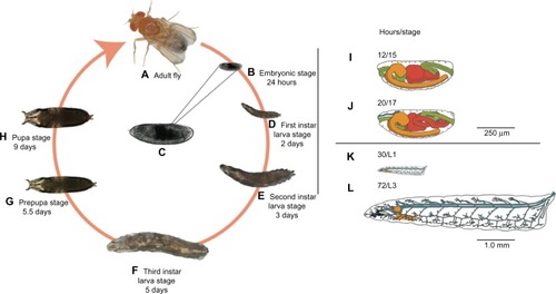
Figure 3 Comparative morphology between nanoparticle-treated and untreated Drosophila embryos.
Notes: Untreated stage 15 embryo (A) is used as reference to determine mortality of embryos that did not progress past stage 15 after delivery of nanomaterials (B). During late embryogenesis (C), rhythmic muscle contractions and a gas-filled tracheal system (arrowhead) are prominent developmental hallmarks. We used the absence of muscle contractions in the presence of the gas-filled tracheal system to determine (D) survival after initial nanoparticle delivery and failure to progress to the first instar (L1) wandering larval stages (E). Mortality at the L1 stage (F) was characterized by a fully developed tracheal system and mouth hooks by fully developed L1 development but failed to progress to later developmental stages. These individuals showed a developed tracheal system and mouth hooks (arrows in E), but no locomotion and no visceral muscle contractions. Scale bars =140 μm.
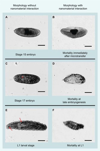
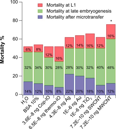
Figure 4 Mortality of Drosophila embryos after microtransfer of nanomaterials at predicted environmental concentrations.
Notes: The effects of the nanomaterials were compared with the effects caused by microtransferring the liquid in which these were diluted. Treatments with IO nanoparticles are compared with treatment with H2O (grey background), and Ag, Au, TiO2, SWCNT, and MWCNT are compared with BSA 10% (white background). For each condition tested, n=50. *Statistical relevance was established through Fisher’s exact test (P<0.005).
Abbreviations: BSA, bovine serum albumin; IO, iron oxide; SWCNT, single-wall carbon nanotube; MWCNT, multiwall carbon nanotube; Ag, silver; Au, gold; TiO2, titanium dioxide; H2O, water; Cop, coprecipitation; Thermo, thermal decomposition.