Figures & data
Figure 1 Deficiency of retroplacental sonolucent zone.
Note: Image courtesy of the Department of Diagnostic Radiology, Queen Mary Hospital, Hong Kong.
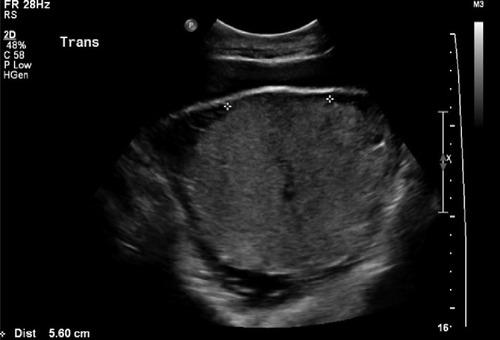
Figure 2 Vascular lacunae.
Note: Image courtesy of the Department of Diagnostic Radiology, Queen Mary Hospital, Hong Kong.
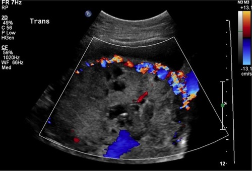
Figure 3 Myometrial thinning.
Note: Image courtesy of the Department of Diagnostic Radiology, Queen Mary Hospital, Hong Kong.
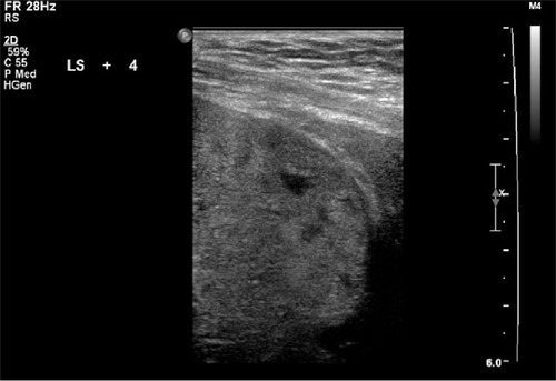
Figure 4 Interruption of bladder line.
Note: Image courtesy of the Department of Diagnostic Radiology, Queen Mary Hospital, Hong Kong.
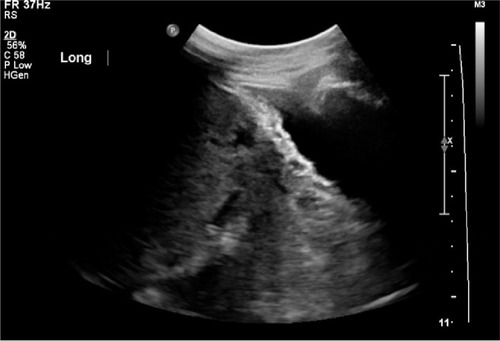
Table 1 Summary of sonographic features of placenta accrete
Figure 5 Ultrasound examination with Doppler study showing a major anterior placenta previa accreta with bladder wall involvement.
Notes: (A) and (B) -Transvaginal ultrasound scan images revealed no myometrial tissue between the lower uterine wall and the bladder. An abnormal vessel running within the bladder wall can also been seen (white arrows). (C) and (D) - Coronal and sagittal magnetic resonance images indicated a bulge at the bladder wall (black arrowheads). Image courtesy of the Department of Diagnostic Radiology, Queen Mary Hospital, Hong Kong.
Abbreviation: MRI, magnetic resonance imaging.
Abbreviation: MRI, magnetic resonance imaging.
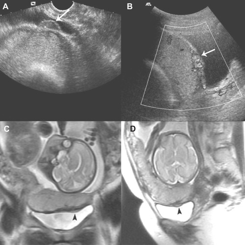
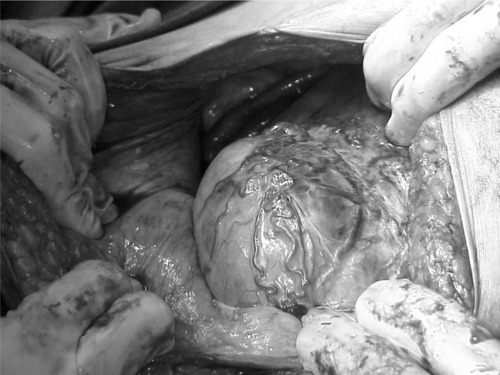
Figure 6 Thin and very vascular uterine lower segment at the time of cesarean section.
Note: The rich vascularity usually correlates with the position of placentation.