Figures & data
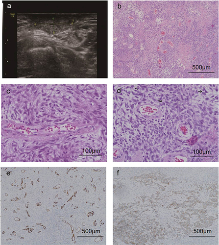
Figure 1 Ultrasonography and pathology of AMFB. (A) Ultrasound indicates a moderate echo in the mass. (B) The hypercellular areas of cells are alternately distributed with hypocellular areas of cells, and abundant thin-walled small blood vessels are seen (HE×100). (C) The hypercellular areas of cells, tumor cells are distributed in bundles or patches around blood vessels, cells are spindle-shaped, cytoplasmic eosinophilic, and some cells are round or oval and epithelioid in shape (HE×400). (D) One case has slightly abundant tumor cells, and the mitotic phase is visible (about 1–2/10HPF)(HE×400). (E) Vascular endothelial cells CD34(+)(IHC×100). (F) Tumor cell vimentin(+)(IHC×100).