
Figures & data
Figure 1 Wnt signaling pathways and the biology of sclerostin.
Notes: (A) Canonical Wnt signaling: in the absence of sclerostin, Wnt binds to LRP 5/6 and its co-receptor, frizzled. This results in phosphorylation of the cytoplasmic tail of LRP 5/6, which allows axin to bind the receptor complex Axin binding leads to inhibition of GSK-3β, which normally functions to target β-catenin for degradation. Therefore, cytoplasmic levels of β-catenin increase and are translocated to the nucleus, where they bind to DNA binding proteins and activate target gene promoters. This results in osteoblast differentiation, proliferation and survival and hence, increased bone formation. (B and C) Loss-of-function of LRP5 and Wnt prevent canonical Wnt signaling: Loss-of-function of LRP5 and Wnt prevent formation of the active Wnt-LRP 5/6-frizzled complex and prevent Wnt signaling. The cytoplasmic tail of LRP 5/6 remains unphosphorylated. Therefore, axin does not bind the receptor complex. GSK-3β activity is uninhibited and therefore leads to phosphorylation of β-catenin, targeting it for degradation. Cytoplasmic levels of β-catenin decrease. Therefore, there is less translocation of the protein to the nucleus. Target gene promoters of the Wnt signaling pathway are not activated. This results in decreased bone formation and increased bone resorption and hence, skeletal fragility and fractures. (D) Inhibition of canonical Wnt signaling by sclerostin: sclerostin is secreted by osteocytes. It binds to LRP 5/6, which prevents Wnt from binding to LRP 5/6 and its co-receptor, frizzled. Therefore, Wnt signaling is inhibited. Through the mechanisms described above (B and C), this results in decreased bone formation and increased bone resorption.
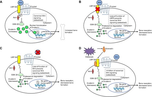
Figure 2 Clinical effects of sclerosteosis and van Buchem disease.
Notes: (A) Sclerosteosis: facial features of a patient with sclerosteosis including a high forehead and large protruding chin. Reprinted from: The American Journal of Human Genetics, 64(6), Balemans W, van Den ende J, Freire Paes-Alves A, et al. Localization of the gene for sclerosteosis to the van Buchem disease-gene region on chromosome 17q12-q21. 1661–1669, Copyright © 1999; with permission from Elsevier.Citation66 (B) van Buchem disease: X-rays show the generalized sclerosis seen in van Buchem patients. The picture to the left is a lateral view of the elbow showing diffuse diaphyseal sclerosis with a thickened cortex. The picture in the center and to the right are anteroposterior and lateral views of the skull which show extensive sclerosis of the calvarium and the skull base, enlargement of the mandible, and obliteration of the paranasal and mastoid air spaces. Reprinted from The American Journal of Human Genetics. 62(2). van Hul W, Balemans W, van Hul E, et al. van Buchem disease (hyperostosis corticalis generalisata) maps to chromosome 17q12-q21. 391–399. Copyright © 1998; with permission from Elsevier.Citation28
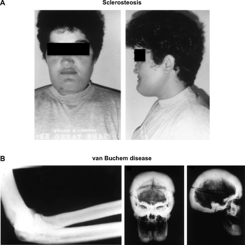
Figure 3 The effect of sclerostin inhibition on Wnt signaling.
Notes: Sclerostin is secreted by the osteocyte. Romosozumab, a humanized MAb against sclerostin, binds circulating sclerostin. This prevents binding of sclerostin to LRP 5/6. Therefore, Wnt is able to bind LRP 5/6 and its co-receptor, frizzled. This activates the Wnt signaling pathway, which eventually leads to osteoblast differentiation, proliferation and survival and, hence, increased bone formation.
Abbreviation: MAb, monoclonal antibody.
Abbreviation: MAb, monoclonal antibody.
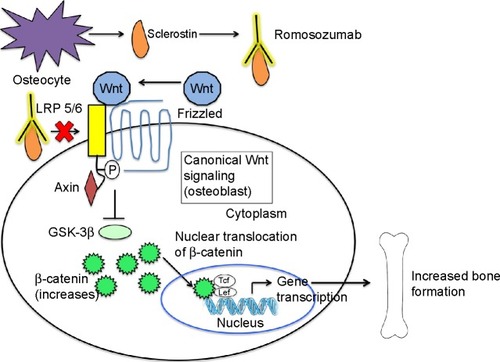
Table 1 Summary of Phase I and II studies of romosozumab
Table 2 Summary of Phase III studies of romosozumab in osteoporosisTable Footnote*