Figures & data
Figure 1 Schematic drawing depicting the domain structure of MMPs.
Notes: The signal peptide guides the MMP through the rough endoplasmic reticulum during synthesis and is cleaved off during secretion by the docking enzyme; the SH propeptide domain maintains the enzyme inactive by blocking the catalytic site, and it is removed or unfolded for MMP activation; the catalytic domain contains the active site of the enzyme and the Zn2+-binding segment. This basic structure is contained in MMP-7 and MMP-26 (matrilysins). MMP-2 and MMP-9 contain three fibronectin-like type II repeats between the active site and the Zn2+-binding segment, and these are responsible for the gelatin-binding property. MMP-9 contains an additional O-glycosylated region, which confers flexibility to the molecule. Except for MMP-7 and MMP-26, all other MMPs contain a carboxy-terminal hemopexin domain, which confers specificity and interacts with many ligands and receptors. The hemopexin and catalytic domains are connected by a small hinge region. MT-MMPs have an additional transmembrane domain and a short cytoplasmic tail or a GPI linkage, which anchor MT-MMPs to the cell membrane.
Abbreviations: MMP, matrix metalloproteinase; SH, sulfhydryl-containing; GPI, glycosylphosphatidylinositol; MT, membrane-type.
Abbreviations: MMP, matrix metalloproteinase; SH, sulfhydryl-containing; GPI, glycosylphosphatidylinositol; MT, membrane-type.
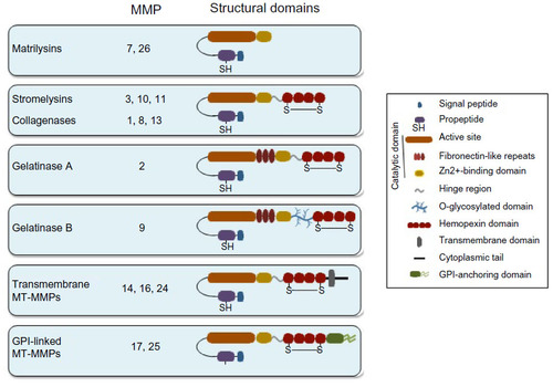
Table 1 Molecular interactions and biological effects involving noncatalytic MMP domains
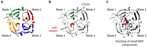
Figure 2 Ribbon diagram of the monomeric hemopexin domain of MMP-9 (PEX9) showing the location of drug target sites.
Notes: (A) PEX9, similar to other PEX domains of MMPs, is composed of four structural blades surrounding a central cavity. (B) Spatial location of the P3 and P6 amino acid sequences within PEX9 (research originally published in The Journal of Biological Chemistry. Ugarte-Berzal E, Bailón E, Amigo-Jiménez I, Albar JP, García-Marco JA, García-Pardo A. A novel CD44-binding peptide from the pro-matrix metalloproteinase-9 hemopexin domain impairs adhesion and migration of chronic lymphocytic leukemia (CLL) cells. The Journal of Biological Chemistry. 2014; 289(22):15340–15349. © the American Society for Biochemistry and Molecular Biology.Citation79). P3 is located in blade 4 and interacts with α4β1 integrin; P6 is located in blade 1 and binds to CD44.Citation79 P3 and P6 act synergistically to inhibit MMP-9-induced CLL cell adhesion and migration, thus serving as therapeutic peptides and simultaneously pointing to “druggable” targets in PEX9. An additional CD44-binding sequence located in the outermost strand of blade 1 has also been reported. (C) Diagram showing the binding sites within PEX9 of small MW compounds mapped by in silico docking (adapted from Cancer Research, Copyright 2011;71(14):4977–4988. Dufour A, Sampson NS, Li J, et al. Small-molecule anticancer compounds selectively target the hemopexin domain of matrix metalloproteinase-9, with permission from AACRCitation82). As observed, both the small compounds and the P3 and P6 sequences are located in close proximity within the central cavity of PEX9. Targeting this region may therefore be a useful approach to control the pathogenic functions of MMP-9, particularly in cancer and inflammation.
Abbreviations: MW, molecular weight; MMP, matrix metalloproteinase; PEX, hemopexin domain; CLL, chronic lymphocytic leukemia.
Abbreviations: MW, molecular weight; MMP, matrix metalloproteinase; PEX, hemopexin domain; CLL, chronic lymphocytic leukemia.