Figures & data
Table 1 Definitions of all types of lesions and infiltrations
Table 2 MRI findings in SCNSL
Figure 1 Common MRI appearance of SCNSL in our group.
Notes: Multiple lesions in brain parenchyma with intermediate intensity on T2-weighted images (A; arrows), with restricted diffusion on diffusion-weighted imaging (B; arrows) and ADC map (C; arrows). Visible vasogenic edema around the lesions on FLAIR (D; arrows). Strong enhancement is present after gadolinium injection on T1-weighted images (E and F; arrows).
Abbreviations: ADC, apparent diffusion coefficient; FLAIR, fluid-attenuated inversion recovery; MRI, magnetic resonance imaging; SCNSL, secondary central nervous system lymphoma.
Abbreviations: ADC, apparent diffusion coefficient; FLAIR, fluid-attenuated inversion recovery; MRI, magnetic resonance imaging; SCNSL, secondary central nervous system lymphoma.
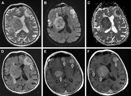
Figure 2 SCNSL with hypothalamic–hypophyseal axis involvement.
Notes: Hypothalamus (white arrow) and optic tracts (black arrows) involvement is visible on T2-weighted images (A) and FLAIR (B), with restricted diffusion in the hypothalamic lesion (C; arrow). The hypothalamic lesion was strongly enhanced after gadolinium injection compared to native T1-weighted images (D; arrow) and post-contrast T1-weighted images (E; arrow). Sagittal post-contrast T1-weighted images (F) show both hypothalamic (black arrow) and hypophyseal involvement with infundibular thickening (white arrow).
Abbreviations: FLAIR, fluid-attenuated inversion recovery; SCNSL, secondary central nervous system lymphoma.
Abbreviations: FLAIR, fluid-attenuated inversion recovery; SCNSL, secondary central nervous system lymphoma.
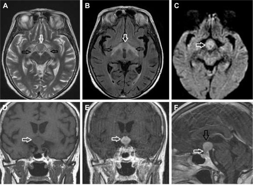
Figure 3 SCNSL mimicking multiple ischemic lesions.
Notes: Multiple hyperintense lesions on T2-weighted images (A) and FLAIR (B) were observed (white arrowheads). The lesions showed diffusion restriction (D; white arrow) and low signal on the ADC map (E; white arrow). Nearly all lesions had signs of hemorrhage on the gradient echo sequence (C and F; black arrows).
Abbreviations: ADC, apparent diffusion coefficient; FLAIR, fluid-attenuated inversion recovery; SCNSL, secondary central nervous system lymphoma.
Abbreviations: ADC, apparent diffusion coefficient; FLAIR, fluid-attenuated inversion recovery; SCNSL, secondary central nervous system lymphoma.
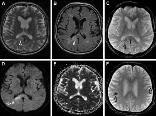
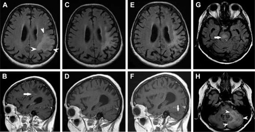
Figure 4 SCNSL mimicking PML.
Notes: Initial MRI at the time of new neurological symptoms showed a single hyperintense lesion on FLAIR (A; arrowheads) with subtle worm-like inhomogeneous enhancement after gadolinium injection on T1-weighted images (B; arrow). On follow-up MRI 7 months later (C and D) the brain lesion decreased in size, and post-contrast enhancement on T1-weighted images completely regressed. The final MRI 4 months later at the time of clinical relapse (E–H) demonstrated that the initial lesion had not changed (E); however, multiple new infiltrative lesions appeared hyperintense on FLAIR, primarily in the brain stem (G; arrow) and cerebellum (H; arrowheads), without post-contrast enhancement on T1-weighted images (F; arrow).
Abbreviations: FLAIR, fluid-attenuated inversion recovery; MRI, magnetic resonance imaging; PML, progressive multifocal leukoencephalopathy; SCNSL, secondary central nervous system lymphoma.
Abbreviations: FLAIR, fluid-attenuated inversion recovery; MRI, magnetic resonance imaging; PML, progressive multifocal leukoencephalopathy; SCNSL, secondary central nervous system lymphoma.