
Figures & data
Figure 1 Hypothesized mechanism of action of near-infrared light (NIR) photobiomodulation.
Notes: NIR (600–980 nm) penetrates tissue to variable depth depending on wavelength, coherence, time, and the tissue involved. A portion of the photonic energy reaches the mitochondria and is absorbed by cytochrome c oxidase. In addition to inducing increased adenosine triphosphate (ATP) production, NIR appears to initiate increased production of reactive oxygen species (ROS), reactive nitrogen species (RNS), and possibly (?) nitric oxide (NO). Downstream events include increased early response genes – c-fos, c-jun – and activation of nuclear factor kappa-B (NF-κB), which in turn induces increased transcription of gene products leading to neurogenesis, synaptogenesis, and increased production of growth factors and inflammatory mediators.
Abbreviation: ↑, increase.
Abbreviation: ↑, increase.
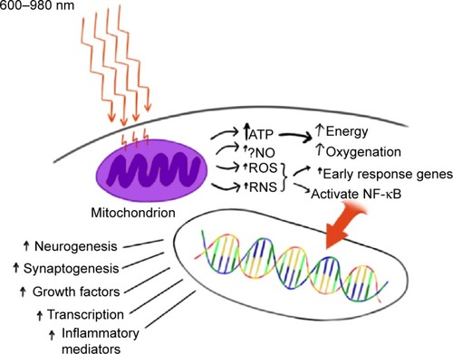
Table 1 Data on infrared light penetration of ex vivo skin samples
Figure 2 Ex vivo human skin studies illustrated.
Notes: (A) The pad of LEDs is held 2 mm from the surface of the light meter detector. The arrow indicates a row of near-infrared light (NIR) LEDs with a wavelength of 880 nm. The meter reads 0.01 W. (B) Human skin 1.9 mm thick is interposed between the NIR LED and the light meter detector. Thin plastic wrap covers the detector. (C) The NIR LED is covered with thin plastic wrap and placed directly against the sample of human skin. Photonic energy could not be detected passing through 1.9 mm of human skin.
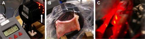
Table 2 Data on infrared light penetration of ex vivo human skin samples
Figure 3 Ex vivo brain tissue studies illustrated.
Notes: (A) The photonic energy penetrating a fixed distance (3 cm) of air was determined. (B) A section of ex vivo lamb head was prepared which included skull, tissue, and brain. (C) The section was interposed in the space between the infrared light emitter and the light meter detector, both of which were fixed in place. The amount of infrared light energy penetrating the fixed distance (3 cm) through tissue was determined. (D) The temperature change was determined using a digital thermometer before and immediately after infrared light exposure.
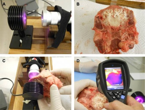
Table 3 Data on infrared light penetration of ex vivo lamb skull, tissue, and brain
Table 4 Data on infrared light penetration of in vivo human tissue