Figures & data
Figure 1 Brain trauma leads to rupture of the intima, media, and adventitia of the blood vessel (A and B), forming an organized hematoma cavity (C). When the hematoma forms outside the arterial wall, it continues to communicate with the injured vessel, thus predisposing it to re-bleeding (D).
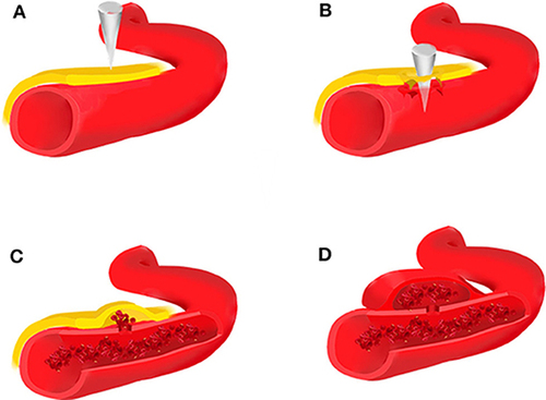
Figure 2 Coronal view of the cavernous sinus including its own contents and the adjacent sphenoidal sinus. This diagram has been reproduced from Gray’s Anatomy 20th US edition which has now lapsed into the public domain.
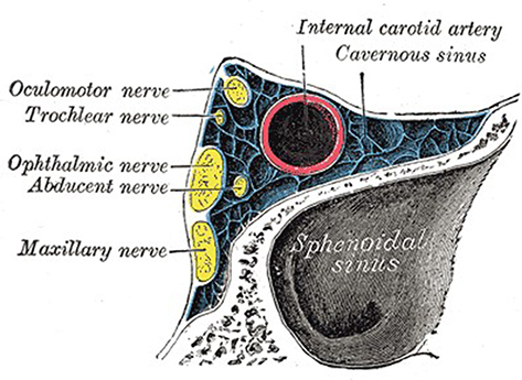
Figure 3 Axial view of the CT showing left-sided extracranial cerebral herniation and retained bullet fragments.
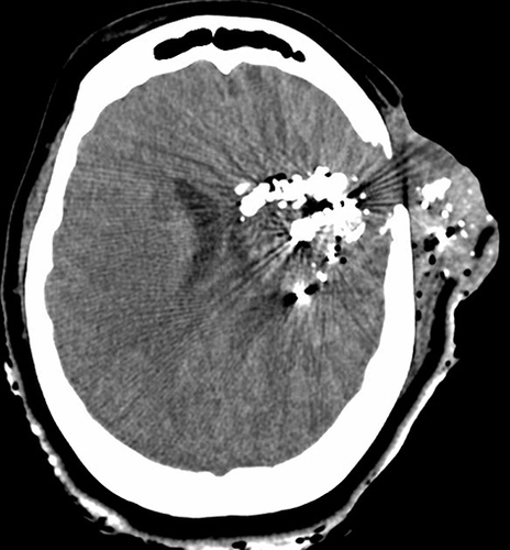
Figure 4 Axial view of the CT showing subarachnoid hemorrhage.
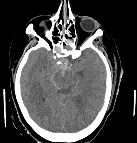
Figure 5 Axial view of the head CT. (Yellow arrow: right-sided lateral sphenoid wall fracture due to force of penetrating trauma).
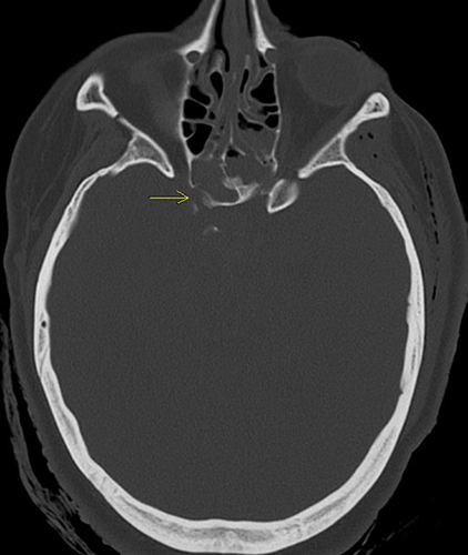
Figure 6 Sagittal view of the head CT. (Yellow arrow: right sided lateral sphenoid wall fracture due to force of penetrating trauma).
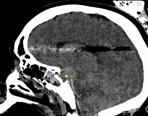
Figure 7 Axial view of the head CT. (Yellow arrow: right sided anterior sphenoid wall fracture due to force of penetrating trauma).
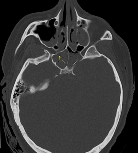
Figure 8 Axial view of the head CT. (Upper arrow: maxillary sinus medial wall fracture due to force of penetrating trauma) (Lower arrow: medial sphenoid septum fracture due to force of penetrating trauma).
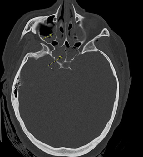
Figure 9 Axial view of the head CT showing VP shunt placement, right to left cerebral midline shift, and external brain herniation.
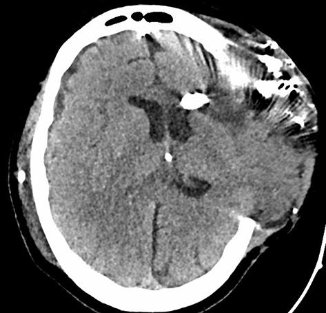
Figure 10 Axial view of the head CT with contrast showing a pseudoaneurysm of the cavernous portion of the right internal carotid artery protruding into the ethmoidal sinus.
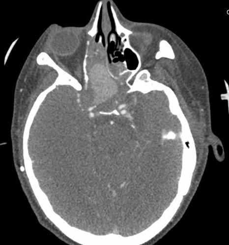
Figure 11 Sagittal view of the head CT with contrast showing a pseudoaneurysm of the cavernous portion of the right internal carotid artery protruding into the ethmoidal sinus.
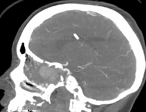
Figure 12 Cerebral angiogram showing large pseudoaneurysm of the cavernous portion of the internal carotid artery.
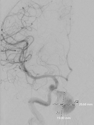
Figure 13 Cerebral angiogram showing ONYX embolization and endovascular coiling of pseudoaneurysm.
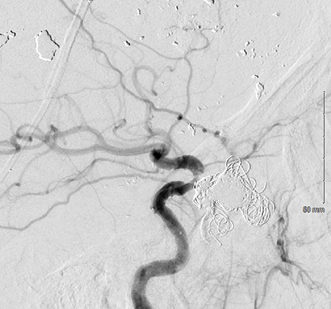
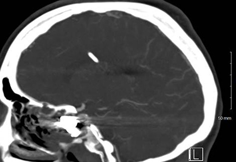
Figure 14 Sagittal view of the head CT showing persistent occlusion of the pseudoaneurysm and internal carotid artery patency.