Figures & data
Figure 1 A confocal image of the eye (stained with phalloidin conjugated with Alexa 488) of Acyrthosiphon pisum depicting the loose arrangement of facets/ommatidia in the eye.
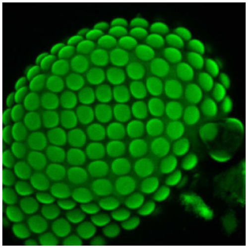
Figure 2 Light micrograph of longitudinally sectioned (LS) light-adapted eye of Acyrthosiphon pisum fixed during the daytime.
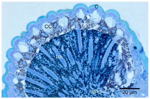
Figure 3 Transmission electron micrograph of sections through the dioptric apparatus of the eye of Acyrthosiphon pisum.
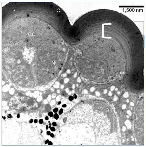
Figure 4 Transmission electron micrograph of sections through the cone of the eye of Acyrthosiphon Pisum.
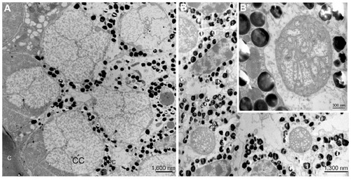
Figure 5 Transmission electron micrograph of the longitudinal section through cornea (C) and cone (CC).
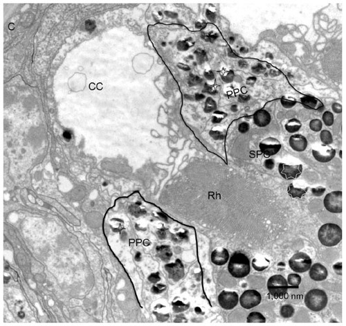
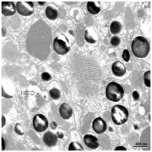
Figure 6 Transmission electron micrograph of the transverse section through the distal rhabdom depicting nuclei (N) of the retinular cells and cone tract (pointed by stars).
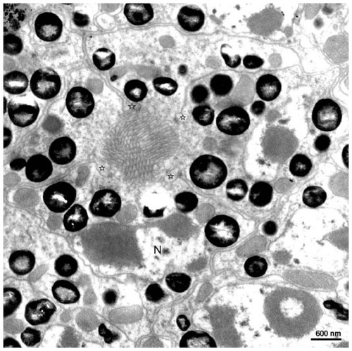
Figure 7 The nuclei of the proximal retinular cells (marked with arrow) and the triangular arrangement of rhabdomere of retinula cell.