
Figures & data
Table 1 Demographic and clinical profile
Table 2 Comparison of results between acute and chronic recurrent Vogt–Koyanagi–Harada (VKH)
Figure 1 (A) A 30-year-old female. BCVA 20/200 in both eyes. Serous retinal detachments, pinpoint leaks, disc hyperflourescence, and pooling of dye. (B) Ultrasonography shows choroidal thickening and serous retinal detachment. (C) OCT – shows large serous retinal detachments with subretinal septae and RPE undulation. (D) Two and a half months after initiating treatment, the retina is reattached. BCVA is RE 20/20 and LE 20/25.
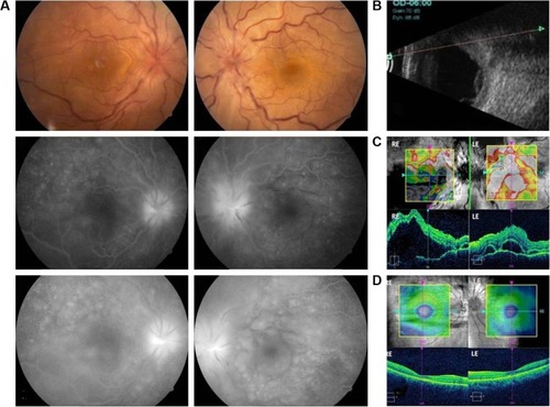
Figure 2 A 25-year-old female with unilateral VKH. (A) RE shows serous retinal detachment and optic nerve head hyperemia. LE fundus is normal. FFA shows RE late leakage and pooling of the dye. (B) RE OCT demonstrates the presence of subretinal fluid and septae, LE OCT is normal. (C) Ultrasonography reveals RE showing a diffuse choroidal thickening and LE showing a normal posterior segment.
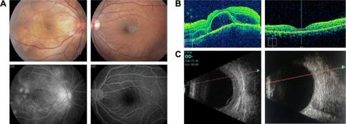
Figure 3 (A and B): Unilateral VKH. A 31-year-old female patient with BCVA RE 20/400 and LE 20/20 shows multiple serous retinal detachments, with multiple pinpoint leaks and subretinal pooling of dye. LE shows normal fundus with normal FFA. (C) Follow-up after 40 months shows both eyes normal fundus with BCVA RE 20/25 and LE 20/20.
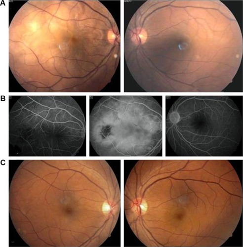
Table 3 Incidence of systemic and ocular manifestations