Figures & data
Figure 1 Retinal images were acquired using the swept-source Deep Range Imaging-OCT (DRI-OCT-1, Topcon, Tokyo, Japan).
Abbreviation: OCT, optical coherence tomography.
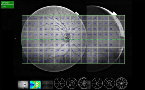
Figure 2 The Deep Range Imaging-OCT segmentation software (Topcon, Tokyo, Japan) was used to identify the limits of the retinal nerve fiber layer as the area expanding from the internal limiting membrane to the inner boundary of the retinal ganglion cell layer.
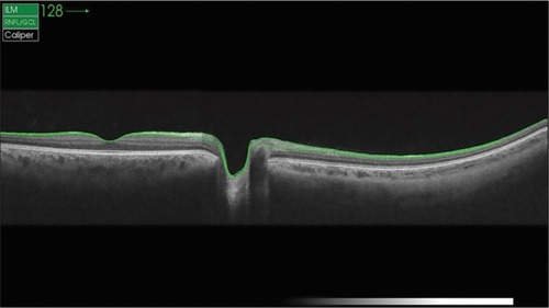
Figure 3 The representative composite images obtained from different age groups containing the retinal nerve fiber layer thickness data: (A) Group 1, (B) Group 2, and (C) Group 3.
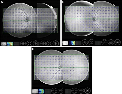
Figure 4 The peripheral RNFL thickness in different age groups.
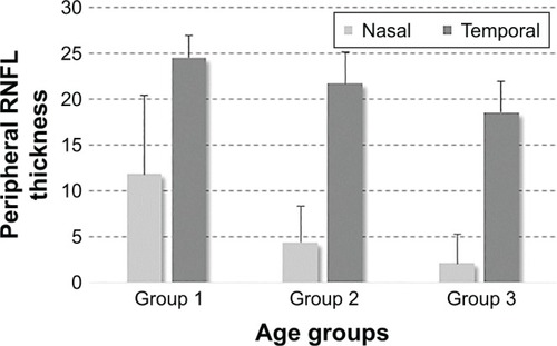
Table 1 The peripheral RNFL thickness
Figure 5 The peripheral-to-peripapillary RNFL thickness ratio in different age groups.
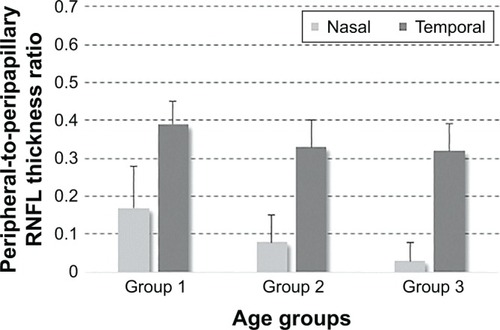
Table 2 The peripheral-to-peripapillary RNFL thickness ratio
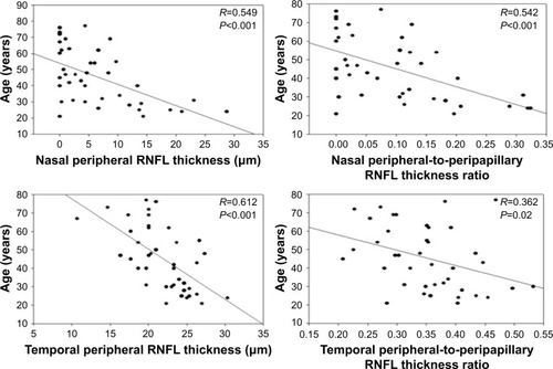
Figure 6 Scatterplots of the relationship between the peripheral RNFL thickness and age.
Abbreviation: RNFL, retinal nerve fiber layer.