Figures & data
Table 1 Patients’ profile of severe Acanthamoeba keratitis cases group and mild Acanthamoeba keratitis cases group
Table 2 Patients’ data at first visit and diagnostic method of severe and mild Acanthamoeba cases
Table 3 Steroid eye drop use before diagnosis, the existence of keratoprecipitates during follow-up, cornea scraping times, visual acuity at last visit, and follow-up period
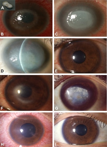
Figure 1 Slit-lamp photographs.
Notes: (A–D) Case S-2. (A) Filthy soft contact lens case, (B) ring ulcer at first visit, (C) disciform infiltrations with keratoprecipitates at 2 months after referral, and (D) corneal scar and mature cataract at 12 months after referral. (E–G) Case S-4. (E) Superficial punctate keratopathy at first visit, (F) ring infiltration with keratoprecipitates at 3 months after referral, and (G) corneal scar at 2 years after referral. (H and I) Case M-5. (H) Keratoneuritis and ciliary injection at first visit, and (I) clear cornea at 3 weeks after referral.