89
Views
0
CrossRef citations to date
0
Altmetric
Original Research

Early Ophthalmic Changes in Macula Does Not Correlate with Visual Function

, , ORCID Icon, , ORCID Icon, ORCID Icon, & show all
Pages 2571-2576 | Published online: 03 Sep 2020

Figures & data

Table 1 Scaling Used to Define Groups with and without Minimal Macular Changes

Table 2 Mean ± SD of Visual Function Tests for 0-A and 0-B Groups

Figure 1 Examples of fundus photos from two study eyes graded as 0-A (left; no minimal macular changes) and 0-B (right; with minimal macular changes). The black circle indicates the region in the macula where fundus retinal changes were observed.

Figure 1 Examples of fundus photos from two study eyes graded as 0-A (left; no minimal macular changes) and 0-B (right; with minimal macular changes). The black circle indicates the region in the macula where fundus retinal changes were observed.

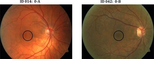
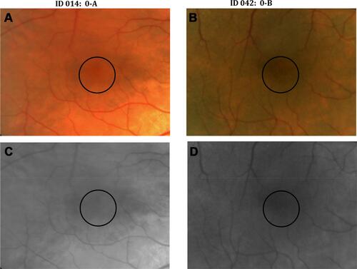
Figure 2 Images from same eyes as shown in under high magnification (A, B) and under gray scale (C, D) to better visualize the minimal macular changes seen in subject ID 042. The black circle indicates the region in the macula where fundus retinal changes were observed.

Figure 2 Images from same eyes as shown in Figure 1 under high magnification (A, B) and under gray scale (C, D) to better visualize the minimal macular changes seen in subject ID 042. The black circle indicates the region in the macula where fundus retinal changes were observed.