
Figures & data
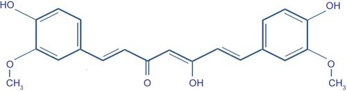
Figure 1 Chemical structure of the curcumin.
Scheme 1 Pathways for ocular disease and biochemical properties of curcumin. Proliferation pathway: CDK4, cyclin D1, c-myc; cell survival pathway: Bcl-2, Bcl-xL; caspase activation pathway: caspase 8/3/9; molecular pathways containing the protein kinase c-Jun N-terminal kinases: JNK; protein kinase B: PKB; reactive oxygen species: ROS; endothelial vascular cell adhesion molecule-1: VCAM-1; intracellular adhesion molecule-1: ICAM-1; leukocyte adhesion molecule-1: ELAM-1; metalloproteinases: MMP; serine protease family: SP; urokinase plasminogen activator system: uPA; tumor suppressor pathway: p53, p21; death receptor pathway: DR4, DR5; cyclooxygenase-2: COX-2; 5-lipoxygenase: 5-LOX; prostaglandin E2: PGE2; nuclear factor –κB: NF-κB; activator protein-1: AP-1; xanthine oxidase: XO; janus kinase, signal transducer and activator of transcription: JAK/STAT; tumor necrosis factor-α: TNF-α; proinflammatory interleukins: IL-1, IL-2, IL-6, IL-8 and IL-12; peroxisome proliferator-activated receptor-γ: PPAR-γ; vascular endothelial growth factor: VEGF; transforming growth factor: TGF-β1; stimulate the fibroblasts expression of fibronectin: FN; collagen.

Figure 2 Spectral domain-optical coherence tomography of a right eye affected by age-related macular degeneration (AMD). Macular neuroepthelial detachment (central thickness of 419 µm) and choroidal neovascular membrane (CNV).
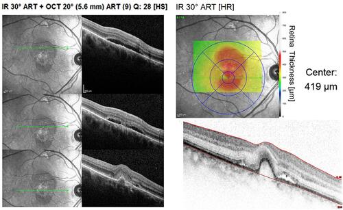
Figure 3 Spectral domain-optical coherence tomography of a right eye affected by diabetic macular edema (ME). Spongy aspect of neuroretin (central thickness of 520 µm) and hyperreflectivity of epiretinal membrane.
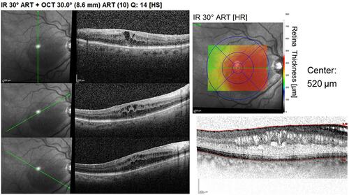
Figure 4 Retinal angiography of a right eye affected by recurrent central serous chorioretinopathy (CSC).
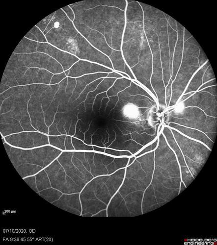
Figure 5 Retinal angiography of a left eye affected by Irvine Gass syndrome. Macular and parapapillary (temporal edge) edema after cataract surgery in a patient suffering from chronic glaucoma with excavated and pale optic disc.
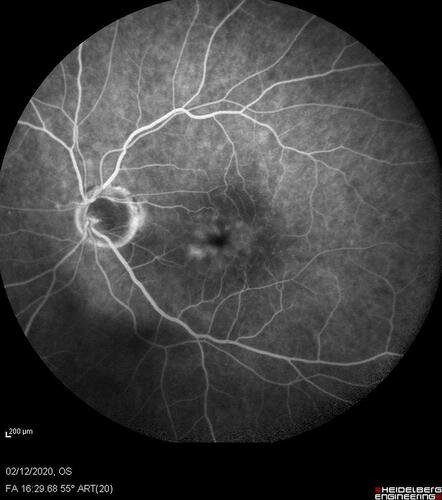
Figure 6 Panels of retinal angiography in central venous thrombosis with ischemic-edema and chorioretinal laser treatment.
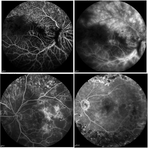
Figure 7 Spectral domain-optical coherence tomography in left eye of retinitis pigmentosa patient. It is possible to observe: an epiretinal membrane between the optic disc and the macula; loss of the photoreceptor layer beyond the fovea; increase of the central nuclear layer.
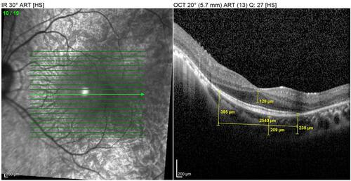
Table 1 Types of Curcumin (Cur) Delivery Systems and Authors (in Bold)