Figures & data
Table 1 Demographic Data
Figure 1 Study methodology: (1) patient selection criteria; (2) data collection; and (3) breakdown of analysis.
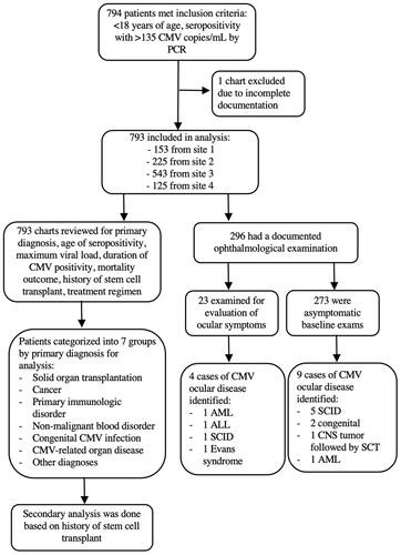
Table 2 Frequency of Clinical Findings and Odds Ratio of Death and Eye Exam by Multivariate Logistic Regression
Table 3 Demographics of Patients with Confirmed CMV Ocular Disease
Figure 2 Examples of fundus findings from our cases of pediatric CMV retinitis. (A) Color photo of left eye with hemorrhagic retinal necrosis (white arrow) and perivascular sheathing (yellow arrow) predominantly involving the macula in a 1-year old male with history of medulloblastoma and autologous hematopoietic progenitor cell transplantation. (B) Optos photo of left eye showing centripetal perivascular retinal whitening (white arrow) in a 15-year-old male with acute lymphocytic leukemia on maintenance chemotherapy of imatinib. (C) Color photos of right eye showing a central white granular lesion of the macula before (white arrow) and 2 months after (Orange arrow) systemic antiviral treatment in a patient with SCID. There was no peripheral involvement in either eye for this patient.
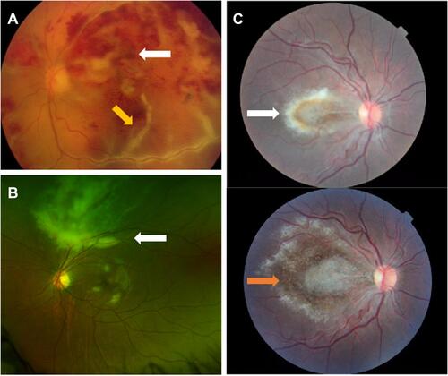
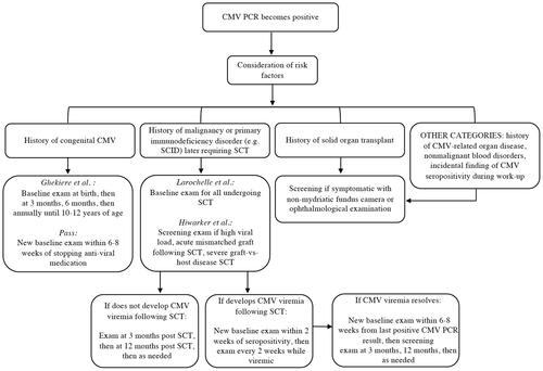
Figure 3 Risk factor-based screening recommendations.