Figures & data
Figure 1 Amyloid deposition appearing as an amorphous, eosinophilic extracellular substance in the stroma of a subepithelial area of the eyelid.
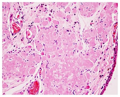
Figure 2 Congo red stain demonstrating orange-red deposits of amyloid.
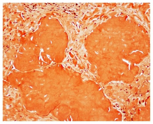
Table 1 Patients’ data and details of periocular and orbital amyloidosis
Figure 3 A 31-year-old woman who presented with a petechial hemorrhage above a conjunctival mass in the inferior area.
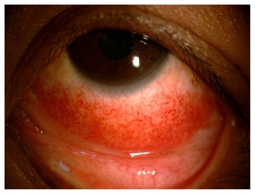
Table 2 Clinical presentation of periocular and orbital amyloidosis