Figures & data
Table 1 Correlation Between LncRNA AZIN1-AS1 Expression and Clinical Features (n=36)
Figure 1 Expression characteristics of AZIN1-AS1 in NSCLC tissues and cell lines. (A) The expression levels of AZIN1-AS1 in cancer tissues and normal tissues was analyzed by GEPIA (TCGA data; LUAD: lung adenocarcinoma). (B) The expression levels of AZIN1-AS1 in normal tissues adjacent to cancer and NSCLC tissues in StarBase database. (C) The expression of AZIN1-AS1 in NSCLC tissues and normal tissues adjacent to cancer was detected by qRT-PCR. (D) The expression of AZIN1-AS1 in normal lung epithelial cell lines BEAS-2B and NSCLC cell lines (HCC-827, NCI-H460, A549 and H1299) was determined by qRT-PCR. *, ***Represent P < 0.05, and P < 0.001 respectively.
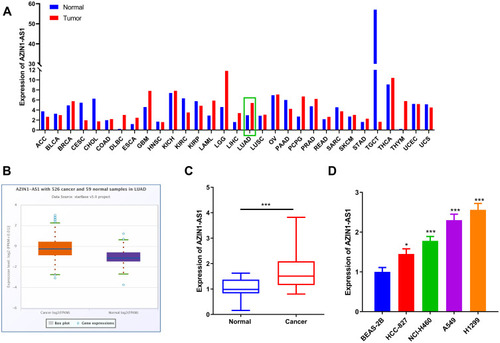
Figure 2 Effects of AZIN1-AS1 on proliferation and metastasis of NSCLC cells in vitro and in vivo. (A) qRT-PCR was used to test the overexpression model of AZIN1-AS1. (B) qRT-PCR was used to test the knockdown model of AZIN1-AS1. (C and D) CCK-8 was used to determine the effects of overexpression and knockdown of AZIN1-AS1 on cell proliferation. (E) EdU assay was used to determine the effects of overexpression and knockdown of AZIN1-AS1 on cell proliferation. (F) Wound healing assay was used to determine the effects of overexpression and knockdown of AZIN1-AS1 on cell migration. (G) Transwell assay was used to determine the effect of overexpression and knockdown of AZIN1-AS1 on the invasive ability. (H) Subcutaneous tumorigenesis test with nude mice was used to determine the effect of knockdown of AZIN1-AS1 on the growth of tumors in vivo. (I) The lung metastasis test was used to determine the metastatic ability of NSCLC cells in mice after knockdown of AZIN1-AS1. *, **, ***Represent P < 0.05, P < 0.01 and P < 0.001 respectively.
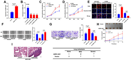
Figure 3 Interaction between AZIN1-AS1 and miR-513b-5p in NSCLC cells. (A) AZIN1-AS1 contained a potential binding site of miR-513b-5p. (B) qRT-PCR was used to detect the expression of miR-513b-5p in normal lung tissues and NSCLC tissues. (C) qRT-PCR was used to detect the expression of miR-513b-5p in normal lung epithelial cell lines and NSCLC cell lines. BEAS-2B was the control group. (D) Correlation analysis was used to analyze the relationship between AZIN1-AS1 and the expression of miR-513b-5p in NSCLC samples. R2=0.411, P<0.001. (E) Luciferase reporter assay was used to detect the binding between miR-513b-5p and AZIN1-AS1-WT. (F) qRT-PCR was used to detect the expression of miR-513b-5p after AZIN1-AS1 overexpression and AZIN1-AS1 knockdown, respectively. **, ***Represent P < 0.01 and P < 0.001 respectively.
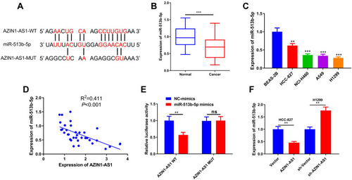
Figure 4 Effects of miR-513b-5p on proliferation and metastasis of NSCLC cells in vitro. (A) qRT-PCR was used to determine the transfection of miR-513b-5p mimics into H1299 cell line. (B) qRT-PCR was used to determine the transfection of miR-513b-5p inhibitors into HCC-827 cell line. (C and D) CCK-8 was used to determine the effects of miR-513b-5p mimics and inhibitors on the proliferation of NSCLC cells. (E) EdU assay was used to determine the effects of effects of miR-513b-5p mimics and inhibitors on the proliferation of NSCLC cells. (F) Wound healing assay was used to determine the migration of NSCLC cells transfected with miR-513b-5p mimics and inhibitors. (G) Transwell assay was used to determine the effects of miR-513b-5p mimics and inhibitors on the invasion of NSCLC cells. *, **, ***Represent P < 0.05, P < 0.01 and P < 0.001 respectively.
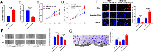
Figure 5 DUSP11 was a target of miR-513b-5p in NSCLC cells. (A) miR-513b-5p contained a potential binding site with the 3ʹUTR of DUSP11. (B) qRT-PCR was used to detect the expression of DUSP11 in normal lung epithelium and NSCLC tissues. (C) qRT-PCR was used to detect the expression of DUSP11 in normal lung epithelial cell lines and NSCLC cell lines. BEAS-2B was the control group. (D) Dual-luciferase reporter assay was used to determine the binding of miR-513b-5p to DUSP11. (E) Correlation analysis was used to analyze the relationship between the expression of miR-513b-5p and DUSP11. R2=0.555, P<0.001. (F) Correlation analysis was used to analyze the relationship between AZIN1-AS1 and DUSP11 expression. R2=0.412, P<0.001. (G) Western blot was used to detect the expression of DUSP11 in NSCLC cells after overexpression and knockdown of AZIN1-AS1. (H) Western blot was used to detect the expression of DUSP11 in NSCLC cells transfected with mimics or inhibitors of miR-513b-5p.**, ***Represent P < 0.01 and P < 0.001 respectively.
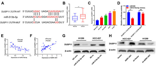
Figure 6 The tumor-suppressive function of miR-513b-5p was partly dependent on its regulatory function on DUSP11. (A) MiR-513b-5p and DUSP11 overexpression plasmids were co-transfected into H1299 cell lines, and the expression of miR-513b-5p in H1299 cells of each group was detected by qRT-PCR. (B) The expression of DUSP11 in H1299 cells was examined by Western blot. (C) CCK-8 method was used to detect the proliferation of H1299 cells after the transfection. (D) EdU assay was performed to examine the proliferation of H1299 cells after the transfection. (E) Wound healing assay was used to detect the migration of H1299 cells. (F) The invasion of H1299 cells was detected by Transwell assay. *, ***Represent P < 0.05, and P < 0.001 respectively. In figure C, &Represents P < 0.05 vs miR-513b-5p mimics group.
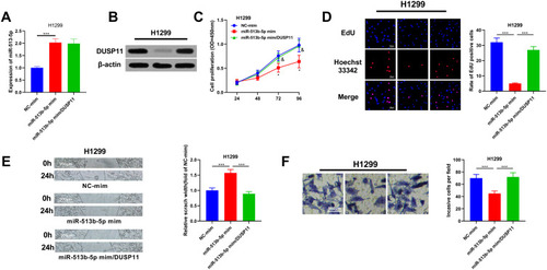
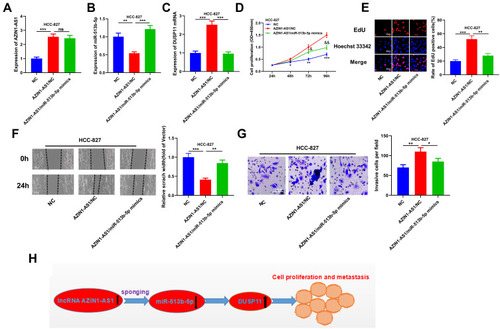
Figure 7 AZIN1-AS1/miR-513b-5p/DUSP11 axis modulated the progression of NSCLC. (A) The level of AZIN1-AS1 in cells co-transfected with miR-513b-5p mimics and AZIN1-AS1 overexpression plasmids was detected by qRT-PCR. (B) The level of miR-513b-5p in cells co-transfected with miR-513b-5p mimics and AZIN1-AS1 overexpression plasmids was detected by qRT-PCR. (C) qRT-PCR was used to detect the level of DUSP11 mRNA in cells co-transfected with miR-513b-5p mimics and AZIN1-AS1 overexpression plasmids. (D) CCK-8 method was used to detect the proliferation of HCC-827 cells. (E) The proliferation of HCC-827 cells was detected by EdU method. (F) Wound healing assay was used to detect the migration of HCC-827 cells. (G) The invasion of HCC-827 cells was detected by Transwell assay. (H) AZIN1-AS1 inhibited miR-513b-5p and up-regulated DUSP11, thus promoting proliferation and metastasis of NSCLC cells.*, **, ***Represent P < 0.05, P < 0.01 and P < 0.001 respectively. In figure D, & and &&Represent P < 0.05, P < 0.01 respectively, vs AZIN1-AS1/NC group.