Figures & data
Figure 1 Sample from the excisional biopsy showing low-grade myxoma.
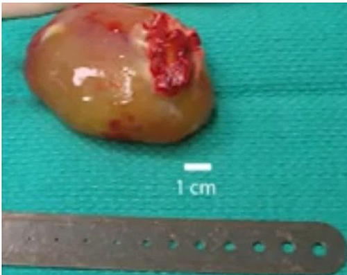
Figure 2 Shows normal electrocardiography of the patient.
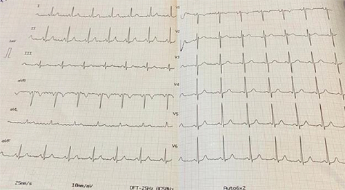
Figure 3 (A) shows the narrow neck of the pseudo-aneurysm with diameter of 12mm (red arrow). (B) showing the pseudo-aneurysm as a mass appearing parallel to the left atrium in this view. (C) longitudinal mass protruding from the pseudo-aneurysm towards the left ventricle. (D) Continues wave Doppler assessment of the prosthetic valve.
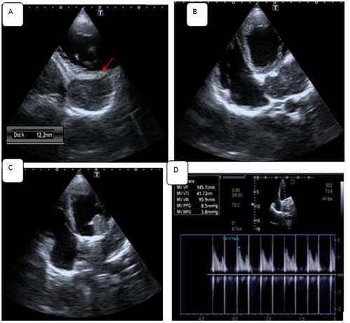
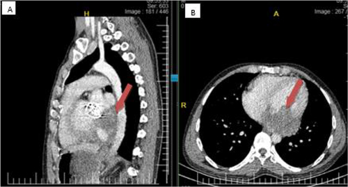
Figure 4 Chest CT -Sagittal (A) and Axial (B) both showing left ventricle showing aneurysmic dilatation and thrombus formation (red arrow).