
Figures & data
Figure 1 Shear wave quality is estimated using a shear wave quality map. Homogeneous green of the left image in the region of interest indicates good quality of shear wave speed estimation; the credibility index is 100%.
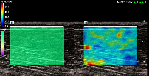
Table 1 Clinical Characteristics of Stroke Patients and Healthy Controls
Table 2 Cross-Sectional Area (CSA) and Thickness Values of BBM Between Stroke Patients (Paretic or Non-Paretic Side) and Healthy Controls
Figure 2 Ultrasound images showed CSA (cross-sectional area) and thickness on the paretic side and non-paretic side of a stroke survivor and a healthy person.
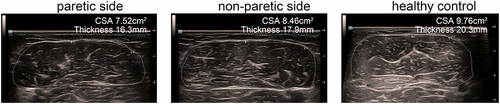
Table 3 Shear Wave Velocity (SWS) Values of BBM Between Stroke Patients (Paretic or Non-Paretic Side) and Healthy Controls
Figure 3 Shear wave elastography of the bilateral biceps brachii muscles (BBM) of stroke patients and healthy individuals, when the upper limbs were positioned at 90° (A) or 45° (B).
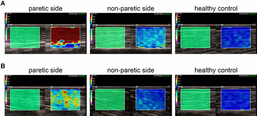
Table 4 SWS Correlation Analysis Using MAS on the Spastic Side