674
Views
33
CrossRef citations to date
0
Altmetric
Endoscopy

Improved detectability of small-bowel lesions via capsule endoscopy with computed virtual chromoendoscopy: A pilot study

, , , , , , , & show all
Pages 1133-1137 | Received 10 Mar 2011, Accepted 25 Apr 2011, Published online: 30 May 2011

Figures & data

Figure 1. Example of an angioectasia. Although some reddening can be seen on the conventional image, the lesion is more clearly visualized by CE-FICE. a: Conventional CE image. b–d: CE-FICE images derived from the three different wavelength settings (b = setting 1; c = setting 2; d = setting 3). CE = conventional endoscopy; CE-FICE = CE with flexible spectral imaging color enhancement.

Figure 1. Example of an angioectasia. Although some reddening can be seen on the conventional image, the lesion is more clearly visualized by CE-FICE. a: Conventional CE image. b–d: CE-FICE images derived from the three different wavelength settings (b = setting 1; c = setting 2; d = setting 3). CE = conventional endoscopy; CE-FICE = CE with flexible spectral imaging color enhancement.

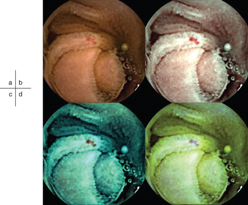
Table I. Number of lesions detected by conventional CE and CE-FICE.

Figure 2. Increase in the number of angioectasias detected per FICE setting (vs. conventional CE). Abbreviations: CE = conventional endoscopy; FICE = flexible spectral imaging color enhancement.

Figure 2. Increase in the number of angioectasias detected per FICE setting (vs. conventional CE). Abbreviations: CE = conventional endoscopy; FICE = flexible spectral imaging color enhancement.

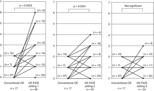
Table II. Capsule video reading times*.