Figures & data
Figure 1. Image enhancement of the anatomical structures allowed with 3D modelling of the visible ear. Top: 3D reconstruction of the middle ear cavities. The ossicular chain appears in blue, the facial nerve in yellow, the cochlea–vestibular apparatus in pink, the carotid artery in red, and the auditory tube in light blue. Bottom: drawings of axial and coronal sections of the middle ear cavities. At least two drawings are required to give information about the 3D arrangement of the structures, whereas the 3D model provides this information directly taking advantage of the 360° rotation to explore several angles of view. Superposition of images or the masking of elements enables the completion or simplification (focussing on, e.g. the auditory ossicles) of the model.
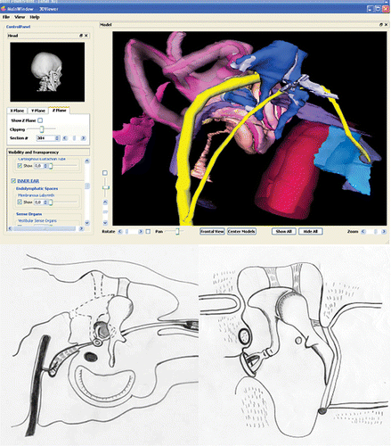
Table 1. Results of the questionnaire filled out by first year undergraduates enrolled on speech therapy and hearing aid practitioner courses
Table 2. Results of the questionnaire filled out by the ENT residents
Figure 2. Comparison of grades in 2007 (lecture only) and 2008 (lecture and use of 3D reconstruction model). For each box, the line represents the median value, the upper point is the 95th percentile and the lower point is the 5th percentile. Error bars are 25th/75th percentile.
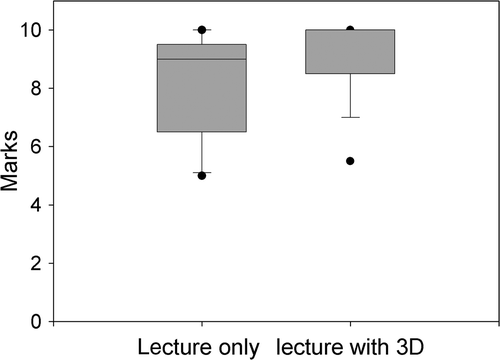
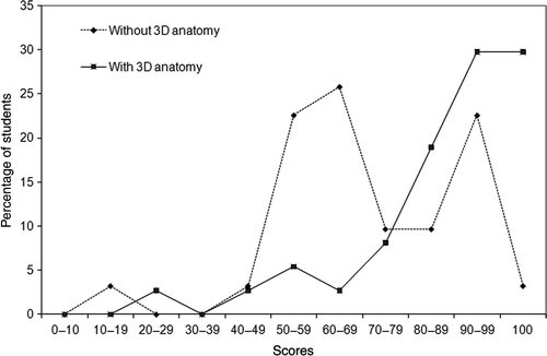
Figure 3. Comparison of marks awarded for test 1 in 2007 (lecture) and those in 2008 (lecture and use of 3D reconstruction model).
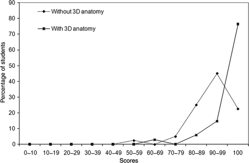
Figure 4. Comparison of marks awarded for test 2 in 2007 (lecture) and those in 2008 (lecture and use of 3D reconstruction model).