Figures & data
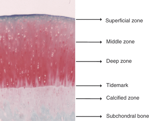
Figure 1. Structure of cartilage. The main difference observed between the chondrocytes in hyaline cartilage is their morphological variation between the zones. Chondrocytes in the superficial zone are flattened and elongated, whereas the cells in the middle zone appear rounded and those in the deep zone have an ellipsoid morphology.

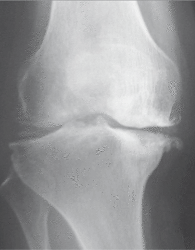
Figure 2. Osteoarthritis of the knee. The knee joint is the most common site of osteoarthritis. The anteroposterior radiograph reveals the valgus deformity of the joint, joint space narrowing, osteophyte formation and sclerosis of subchondral bone. In knee osteoarthritis, medial femorotibial alterations often predominate.

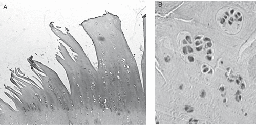
Figure 3. Histological features of cartilage in osteoarthritis. Osteoarthritic cartilage demonstrates severe fibrillation leading to cracking that extends into the tidemark. There is an obvious decrease in the numbers of chondrocytes in the late phase of the disease (A), while cluster formations, corresponding to chondrocyte proliferation, are sometimes observed to be adjacent to the area of fibrillation in the early phase (B).

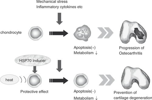
Figure 4. A schematic drawing of the events involved in the initiation and progression of osteoarthritis. Potential causative factors are listed above and the cellular and morphological changes are listed below Citation[12]. Articular cartilage is an important component of the joint, and it is always exposed to pressure produced by weight bearing and muscle contraction. If the mechanical stress increased to a level excessively higher than the physiological levels, the cartilage matrix will be impaired and osteoarthritis could occur. In addition to mechanical stress, its onset and progression could be induced by inflammatory cytokines produced by chondrocytes in cartilage or the synovial tissue in the joint. Inflammatory cytokines contribute to the dysregulation of the chondrocyte function. The association of increased production of metalloproteinases induced by inflammatory cytokines with cartilage damage has been established. The apoptosis of chondrocytes caused by various types of stress has attracted the attention of researchers as an important etiologic factor in OA progression.
![Figure 4. A schematic drawing of the events involved in the initiation and progression of osteoarthritis. Potential causative factors are listed above and the cellular and morphological changes are listed below Citation[12]. Articular cartilage is an important component of the joint, and it is always exposed to pressure produced by weight bearing and muscle contraction. If the mechanical stress increased to a level excessively higher than the physiological levels, the cartilage matrix will be impaired and osteoarthritis could occur. In addition to mechanical stress, its onset and progression could be induced by inflammatory cytokines produced by chondrocytes in cartilage or the synovial tissue in the joint. Inflammatory cytokines contribute to the dysregulation of the chondrocyte function. The association of increased production of metalloproteinases induced by inflammatory cytokines with cartilage damage has been established. The apoptosis of chondrocytes caused by various types of stress has attracted the attention of researchers as an important etiologic factor in OA progression.](/cms/asset/5813f899-8116-4760-8726-9ce0757b2bc7/ihyt_a_410924_f0004_b.gif)
Figure 5. Strategy for applying hyperthermia to the treatment of OA. Hsp70-induction in chondrocytes by heat stimulation and/or HSP inducer, such as glutamine, MG132, GGA and curcumin can have a therapeutic effect on OA by inhibiting chondrocyte apoptosis as well as increasing the cartilage metabolism.

