Figures & data
Figure 1. Changes in Hsp72 levels in pancreatic tissue after pretreatment with whole-body hyperthermia in rats. (A) Hsp72 expression in rat pancreatic tissue 48 h after WBH treatment at 42 °C or ambient temperature was detected by Western blot. (B) Signal intensities were based on densitometry using an image analyser. All data are expressed as the mean ± standard deviation (SD); * denotes a significant difference compared to the negative control group (p < 0.05).
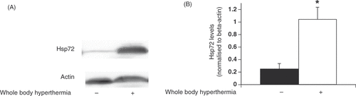
Figure 2. Evaluation of the effects of whole-body hyperthermia on pancreatic tissue in LPS-treated rats by electron microscopy. (A–C) High magnification of beta cell insulin granules. Pancreatic tissue specimens were obtained from the negative control group (A); untreated, LPS group (B); and WBH-treated, LPS group (C). (D) Sections from rats who were administered LPS for 12 h with or without WBH pretreatment. Sections from the three groups were blindly chosen and the number of insulin granules was counted in hyper-magnified fields. All data are expressed as the mean ± SD. # denotes a significant difference compared to the LPS group (p < 0.05); * denotes a significant difference compared to the negative control group (p < 0.05).
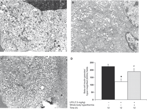
Figure 3. Insulin secretion from pancreatic beta cells of rats with or without whole-body hyperthermia pretreatment and LPS treatment. Insulin secretion from pancreatic beta cells of rats exposed to WBH or ambient temperatures prior to LPS administration. Insulin levels were determined using a rat insulin enzyme-linked immunosorbent assay (ELISA) kit. Harvested cells were incubated with KRB buffer containing the indicated concentrations of glucose. All data are expressed as the mean ± SD. # denotes a significant difference compared to the LPS group (p < 0.05); * denotes a significant difference compared to the negative control group (p < 0.05).
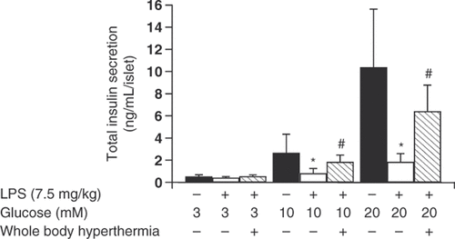
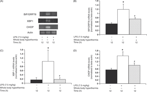
Figure 4. Change in ER stress response gene levels in pancreatic beta cells due to whole-body hyperthermia treatment prior to LPS administration. Rats were treated with LPS following exposure to either 42 °C or ambient temperature. Pancreatic beta cells were harvested and transcripts were analysed by polymerase chain reaction (PCR) and quantified by densitometry. (A) Levels of BiP/GRP78, XBP-1, and CHOP transcripts in cells from LPS-treated rats were determined by RT-PCR. Signal intensities of BiP/GRP78 (B), XBP-1 (C), and CHOP (D) transcripts were based on densitometry using an image analyser. All data are expressed as the mean ± SD. # denotes a significant difference compared to the LPS group (p < 0.05); * denotes a significant difference compared to the negative control group (p < 0.05).