Figures & data
Figure 1. Experimental setup for temperature measurements during laser ablation in human disc specimen. (A) The static main field in the open MRI is vertical (white arrows), the open configuration allows good access from all sides to the scanners' iso-centre. (B) The bare laser fiber (1) and the fiber optic sensor (2) are inserted into the in vitro phantoms. A flexible multi-purpose coil (3) was used for MR-imaging. During heating experiments the dynamically acquired images could be viewed on an in-room monitor (4) next to the scanner. (C) The bare laser fiber and the fiber optic probe are placed orthogonally to one another with a distance of about 4mm inside the disc tissue.
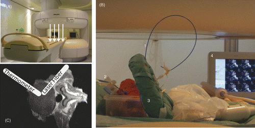
Figure 2. Representative data of the thermal history show the direct temperature measured with the fiber optic probe and the dynamically acquired MRTh data of the unspoiled GRE (TE = 7ms) during the heating experiment. PRF-MRTh with a postprocessing algorithm (•), that subtracts the magnetic field drift (⋄) from the phase-image differences (*), avoids an underestimation of the temperature.
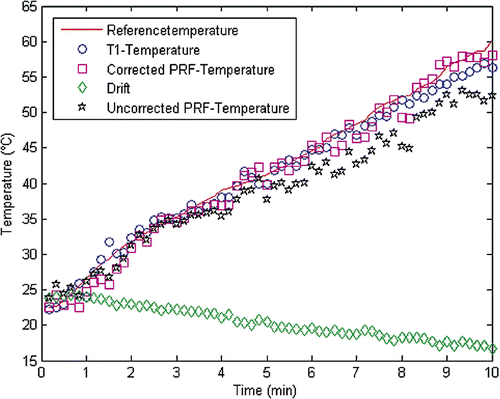
Figure 3. Age-dependency of T2* measured in the nucleus in 20 volunteers (4/group).
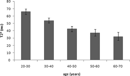
Figure 4. Comparison of four gradient-echo based sequences for thermal mapping. (A) SNR in two different tissues types acquired during volunteer scans, (B) needle susceptibility artifacts in cadaveric lumbar disc tissue.
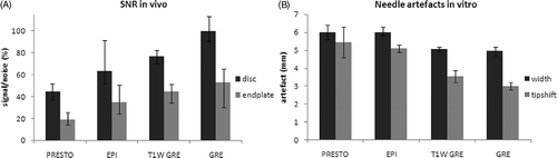
Table I. Results of linear regression analysis of the phantom heating experiments for GRE with T1- and PRF-based MRTh. The analysis was based on a two-parameter model ΔTMR=Tref_min+b · TMR, the number of data points was n = 60 and the temperature range from 20 to 60°C.
Figure 5. Mean-difference plots (Bland-Altman Plots) show the comparison between measured temperature with the optical temperature probe and calculated absolute MR-temperature with a two-parameter model ΔTMR =Tref_min+b · TMR. Results with lowest and highest echo-times for both thermometric methods (PRF, T1) are shown, the means and ±95% limits of agreement are indicated: (A) PRF-thermometry, TE = 2,4ms; (B) T1-thermometry, TE = 2,4ms; (C) PRF-thermometry, TE = 15ms; (D) T1-thermometry, TE = 15ms.
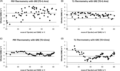
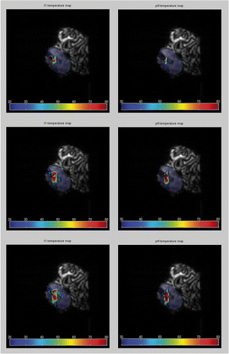
Figure 6. MRTh is calculated from image data during a laser ablation in human cadaveric disc. For imaging the GRE was used (TE = 10ms). Color-coded thermographic plots were mapped over the corresponding MR magnitude images. Calculations based on T1-MRTh (left) and on PRF-MRTh (right) show the heat spread inside the nucleus. The image data were taken at different points during the experiment: (A) after 2 min, (B) after 6min and (C) after 10 min of lasing with a NdYAG laser (1064 nm).