Figures & data
Figure 1. Characterisation of sgrp170. (A) Western blot with polyclonal anti-grp170 on the spent culture media of wild type Colon-26 (lane 1), mock-transfected Colon-26 (lane 2) and sgrp170-transfected Colon-26 (lane 3). (B) The effect of sgrp170 on the inhibition of protein aggregation in vitro. Luciferase alone (filled squares) or with equal molar ratio of sgrp170 (open squares) was incubated at 43°C for 30 min. The light scattering was measured by optical density change at 320 nm every 5 min.
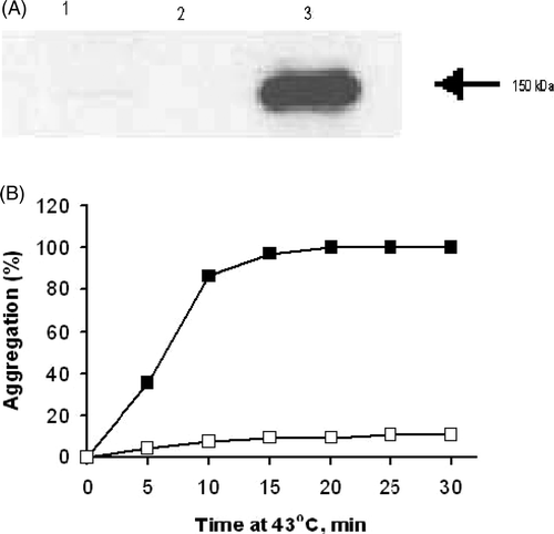
Figure 2. Transfection of Colon-26 with secreted grp170 results in tumour rejection in vivo. Balb/c mice were inoculated with wild type or transfected tumour cells as indicated (n = 5 per group), and the tumour growth measured as described in materials and methods. Tumour growth curves for wild-type Colon-26 (A), mock-transfected Colon-26 (B) and sgrp170-transfected Colon-26 (C) are shown. Growth curves for individual mice are represented by solid lines and the average growth curve is represented by a dotted line.
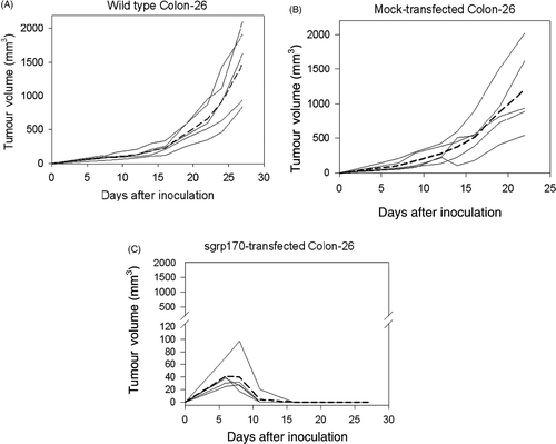
Figure 3. The effect on the growth of sgrp170-transfected Colon-26 tumours by in vivo depletion of both CD8+ and CD4+ T-lymphocytes (A), depletion of the CD8+ subpopulation alone (B) or depletion of the CD4+ subpopulation alone (C). Tumour growth curves for individual mice are represented by solid lines (n = 5 per group), and the average growth curve is represented by the dotted line.

Figure 4. Immunohistochemical localisation of CD3+ immune cells in Colon-26 tumours. On the eighth day of growth, mock-transfected Colon-26 tumour contained a number of CD3 positive immune cells (red arrows) in close proximity to viable tumour cells (blue nuclei) (A). In contrast, after eight days, the site of inoculation with the sgrp170 transfected tumour cells consists of a dense aggregation of CD3 positive immune cells and few viable tumour cells. (20 × original magnification) (B).
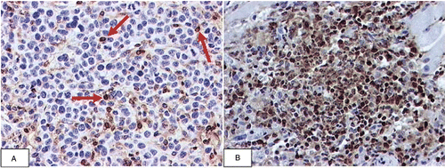
Figure 5. Splenocytes from mice bearing sgrp170 transfected tumours have a significantly higher frequency of AH-1 reactive cells. Splenocytes isolated from naïve BALB/c mice (grey bars), mice bearing wild-type Colon-26 (white bars) or sgrp170-transfected Colon-26 tumours (black bars) were assayed for the frequency of IFN-γ-producing cells by ELISPOT in response to medium alone or the AH-1 epitope of gp70 of MuLV. The error bars indicate the standard error of the measurements from individual mice (n = 3 per group).
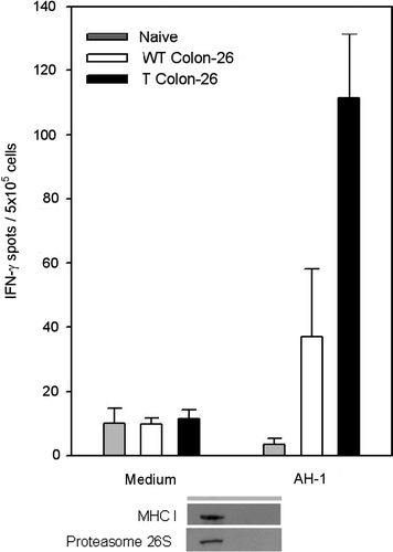
Figure 6. Western blots of the eluants from nickel-chromatography of cell culture supernatants of Colon-26 cells transfected with sgrp170 (T) or mock-transfected Colon 26 (E). Western blot of the T and E fractions using specific antibodies to gp70, gp96, MHC Class 1, proteasome 26S and hsp70 (A). Western blot of the unfractionated culture supernatants of sgrp170-transfected Colon-26 cells (Sup T) and mock-transfected Colon-26 (Sup E) using antibodies specific for gp70 and gp96 (B).
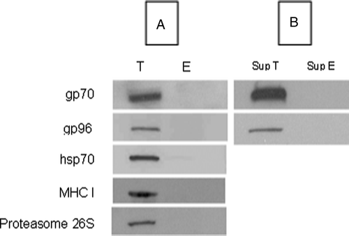
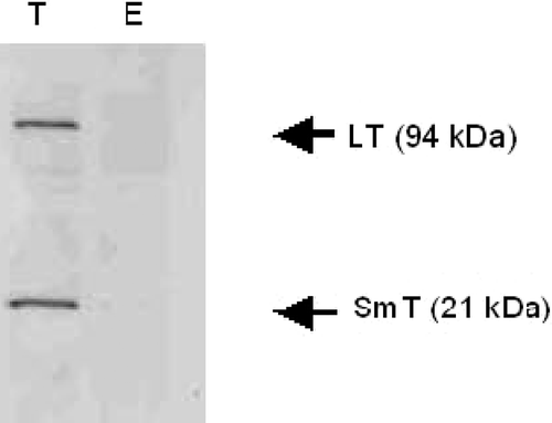
Figure 7. Western blots of the eluants from nickel-chromatography of cell culture supernatants of sgrp170-transfected Cos-7 cells (T) and mock-transfected Cos-7 cells (E) using an antibody specific to both small and large T-antigens.