Figures & data
Figure 1. Typical cases of chest wall recurrence of breast cancer to be addressed with conformal thermotherapy.
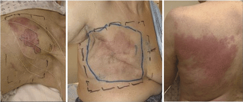
Figure 2. (A) Buried coplanar waveguide feed-line design to excite a single PCB square slot aperture with matching stubs on the fixed-width coplanar slot feeding each excitation port, (B) matched CPW feed-line distribution network to 35 element L-shaped CMA array, and (C) matching 5–10-mm thick water bolus coupling layer with optimised circulation for homogeneous temperature across large surface and integral thermal mapping catheters Citation[34], Citation[35].
![Figure 2. (A) Buried coplanar waveguide feed-line design to excite a single PCB square slot aperture with matching stubs on the fixed-width coplanar slot feeding each excitation port, (B) matched CPW feed-line distribution network to 35 element L-shaped CMA array, and (C) matching 5–10-mm thick water bolus coupling layer with optimised circulation for homogeneous temperature across large surface and integral thermal mapping catheters Citation[34], Citation[35].](/cms/asset/a86c87cf-9ff8-448b-8080-d4fa7232c4a6/ihyt_a_501511_f0002_b.gif)
Figure 3. CMA applicator conformity to contoured chest wall: (A) Photo of 18-element DCC array coupled with 9-mm thick water bolus to torso phantom, (B) CT scan cross section of torso phantom through one row of the 18 element DCC array, (C) Elastic outer support vest, (D) MR scan showing conformal water bolus on a mastectomy patient.
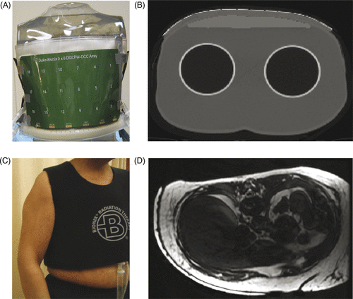
Figure 4. Simulated SAR and heating pattern on contoured surface of an elliptical tissue model. (A) Normalised SAR 10 mm deep in muscle for a 35 element CMA with three centrally located DCC antennas powered off to demonstrate localisation and control of heating; (B) Normalised SAR pattern 10 mm deep in muscle on the contoured torso for a second power configuration as shown inset (selected peripheral apertures in black turned off). (C) Steady-state temperature distribution inside the simulated target volume extending 15 mm deep, as calculated for the SAR distribution of .
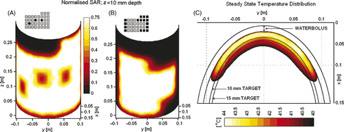
Figure 5. (A) 3 × 6 DCC array (17 × 32 cm) fabricated with three-layer LCP and buried coplanar waveguide feed-lines and matching stubs; (B) measured SAR distribution 10 mm deep in muscle-equivalent phantom for the power configuration with six antennas marked with X's turned off; (C) 50% iso-SAR contour in the 10-mm deep plane for the power configuration shown in ; (D) HFSS simulated SAR pattern for same power configuration tested in ; (E) 3D rendering of HFSS simulation of 25%, 50% and 75% SAR iso-SAR surfaces for same power configuration.
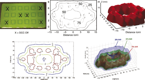
Figure 6. Temperatures measured during clinical hyperthermia with 31 × 14 cm CMA applicator on a chest wall patient. (A) Temperatures measured at 10 mm increments in four catheters on the tumour target surface and one catheter implanted approximately 10 mm deep, prior to adjusting DCC power levels for most uniform heating. (B) Temperatures measured at 10 mm increments along an indwelling catheter 5–10 mm deep in tissue midway through heat treatments of the same patient, once with a 24 aperture conformal array and average of four successive treatments with a 16 element planar array applicator.
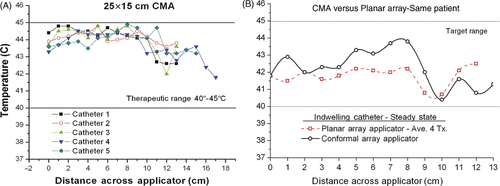
Table I. Same site thermal dosimetry comparison of the best treatments with conformal microwave array (CMA) and planar array (PA) applicators. Negative values mean the planar array produced higher average temperatures for the measured points in target. Total number of measured points in tumour target is higher for CMA, proportionate to the increased treatment area. Note the planar array was limited to treating a portion (169 cm2) of the disease target whereas the CMA coverage ranged from 288 to 500 cm2as needed to cover the entire tumour target.