Figures & data
Figure 1. (a-c) US and MRI scans show abscess formation following microwave ablation (MWA) of intrahepatic cholangiocarcinoma with Cholangiojejunostomy. A 3.2-cm metastasis in a 52-year-old man was treated with MWA. (a) ultrasound scan shows the gas inside bile duct (thin arrows) arrived at ablation zone from liver hilar and hyperechoic ring of gas distribute around ablation zone 3 days after ablation (thick arrow). (b) MRI scan shows the pus developed from ablation zone to right thoracic cavity along needle track and forming a dumbbell-shaped abscess cavity (arrow). (c) Coronal MRI scan shows abscess in right thoracic cavity (thick arrow) coming from ablation zone involved soft tissue of chest wall (thin arrows) along coagulated needle track.
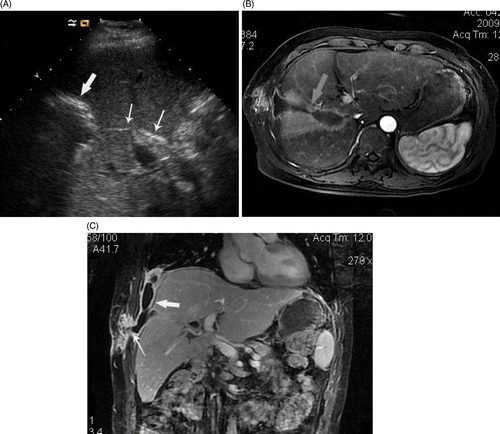
Figure 2. (a, b) MRI and CT scan showed abscess forming after ablation involved right thoracic cavity and inferior lobe of right lung. Four metastasis in a 56-year-old man were treated with MWA. (a) Coronal MRI scan shows two abscess forming 12 days after MWA (thin arrows) and superior abscess involved right thoracic cavity via needle track (thick arrow). (b) transverse CT scan shows pus in right thoracic cavity involved inferior lobe of right lung and induced consolidation of local lung (arrow) and limited inflammatory respond: the patient had symptom of cough and purulent sputum.
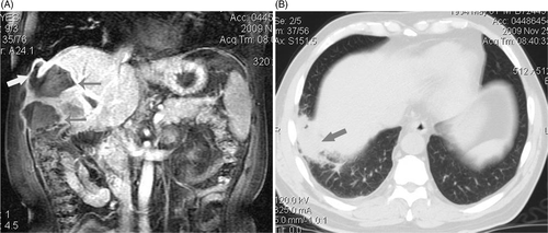
Figure 3. (a-c) transverse CT scan and digital subtraction angiography (DSA) shows abscess formation and a small account of bleeding in abscess cavity. A 53-year-old man had a symptom of bloody stool shortly after MWA of intrahepatic metastasis. (a) Transverse CT scan shows an abscess formation following MRA which had a large fluid- and air-filled cavity (arrow). (b) Hepatic arteriography hadn't found bleeding. (c) Coronal reconstruction of post-arteriography CT shows there was contrast agent deposition inside and around the ablation zone (arrow) in venous phase which help to deduce that there was a small number of active bleeding surrounding ablation zone.
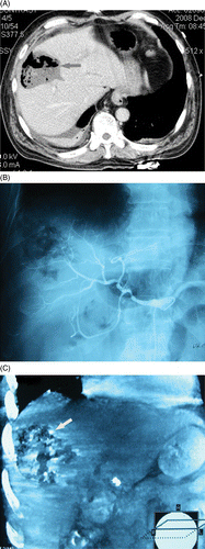
Table I Basic information of liver abscess and management after microwave ablation of liver cholangiocarcinoma metastases in four patients.